X-Ray, CT, US, MRI Phantoms
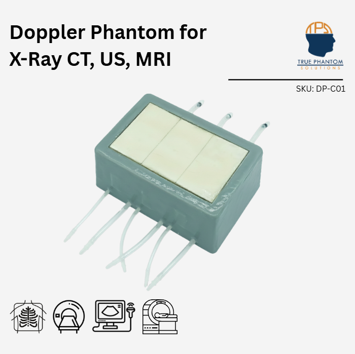
Doppler Phantom for X-Ray CT, US, MRI
$4,442
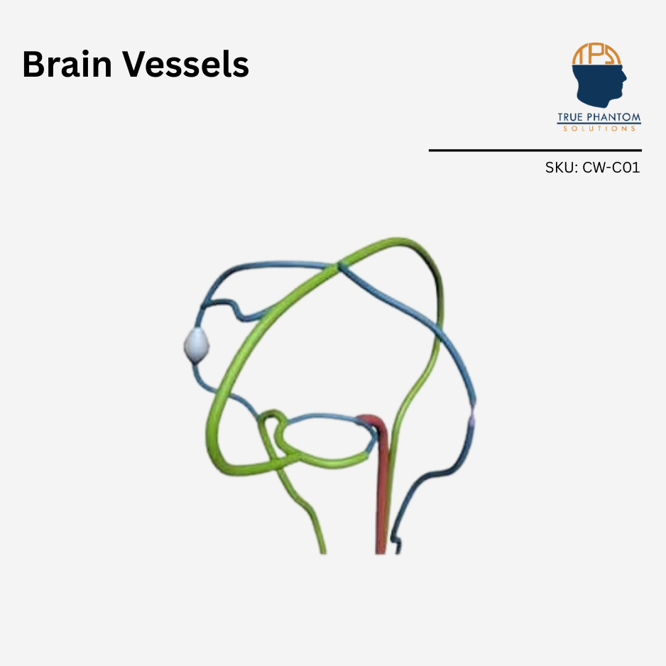
Doppler Phantom serves purposes in medical imaging research, development, training, and testing. The phantom includes a bone plate simulating the 2D projection of the skull with realistic internal porosity. Internal blood vessels can be filled with water or commonly used blood-mimicking fluids
This product can be customized based on your requirements — contact us.
Doppler Phantoms are used for medical imaging research and development, training, and testing purposes. The bone plate installed on the top of this phantom mimics the skull bone, which in this case simulates only the 2D projection of the skull with a realistic internal porosity structure. The internal blood vessels can be filled either with water or with most of the commonly used blood-mimicking fluids. This phantom can be used with Barium-sulfate, Iodine-based, Gadolinium–based, Microbubble, and Iron Oxide-based contrast mediums.
The phantom can be customized in multiple ways to suit the needs of a wide variety of applications.
| Type of Tissue | Sound Velocity[m/s] | Density [g/cm3] | Attenuation Measured at 2.25 MHz [dB/cm] |
| Cortical Bone | 3000 ± 30 | 2.31 | 6.4 ± 0.3 |
| Trabecular Bone | 2800 ± 50 | 2.03 | 21 ± 2 |
| Brain Matter | 1400 ± 10 | 0.99 | 1.0 ± 0.2 |
| Skin Tissue | 1400 ± 10 | 1.02 | 1.7 ± 0.2 |
| HU Values of the Tissue Mimicking Materials | ||
| S.No. | Tissue Type | HU Value (average) |
| 1 | Brain Tissue | -25 |
| 2 | Trabecular Bone | 800 |
| 3 | Cortical Bone | 1300 |
| Section Number | Cortical Outer Thickness [mm] | Trabecular Thickness [mm] | Cortical Inner Thickness [mm] | Total Thickness [mm] |
| A | 1.5 ± 0.2 | 1 ± 0.2 | 1.0 ± 0.2 | 3.5 ± 0.2 |
| B | 1.5 ± 0.2 | 2 ± 0.2 | 1.0 ± 0.2 | 4.5 ± 0.2 |
| C | 2.5 ± 0.2 | 3m± 0.2 | 1.0 ± 0.2 | 6.5 ± 0.2 |
Similar products