
X-Ray, CT, US, MRI Phantoms
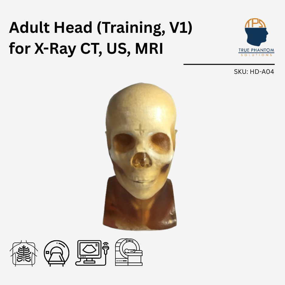
Adult Head (Training, V1) for X-Ray CT, US, MRI
$10,200
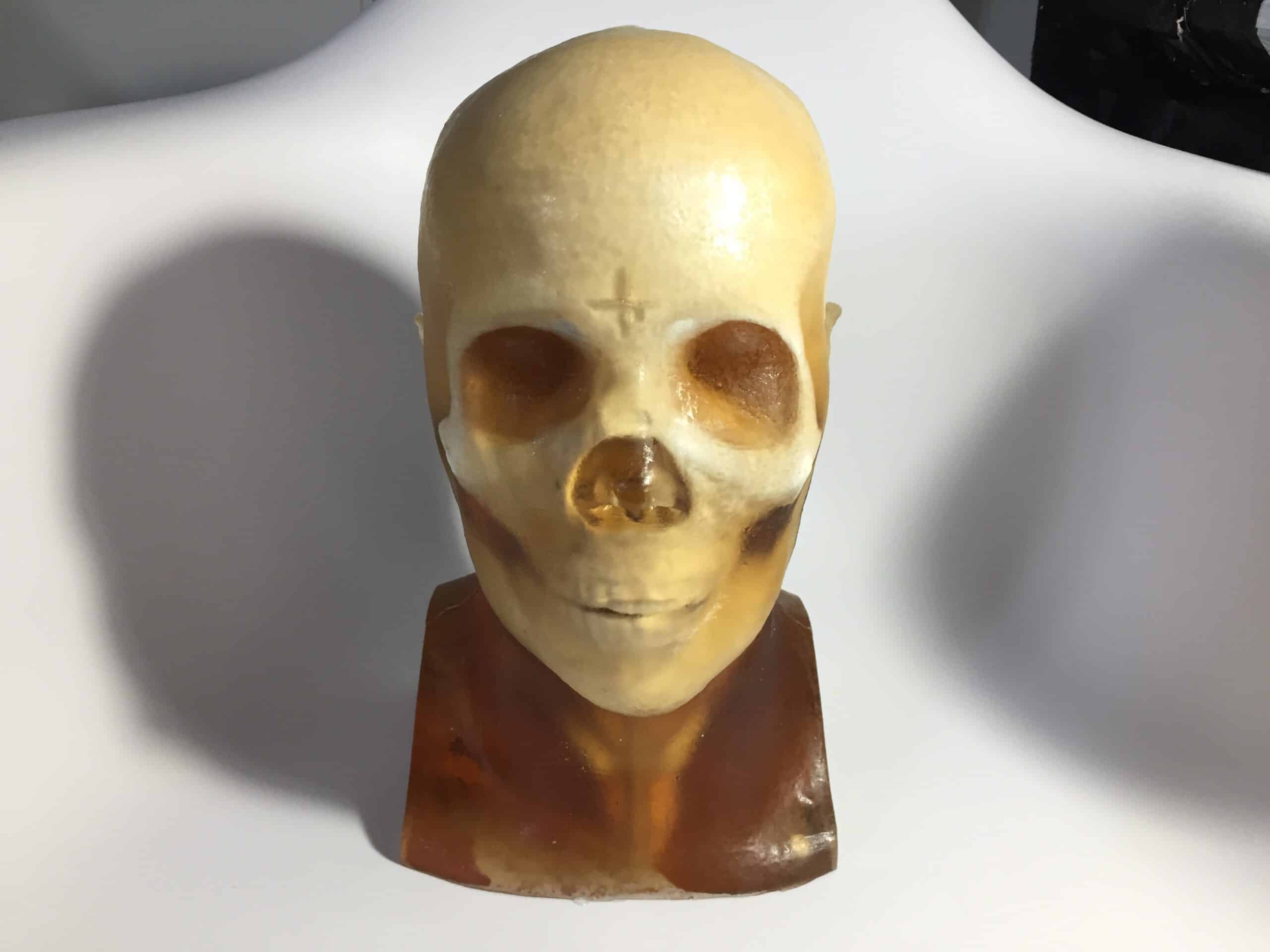
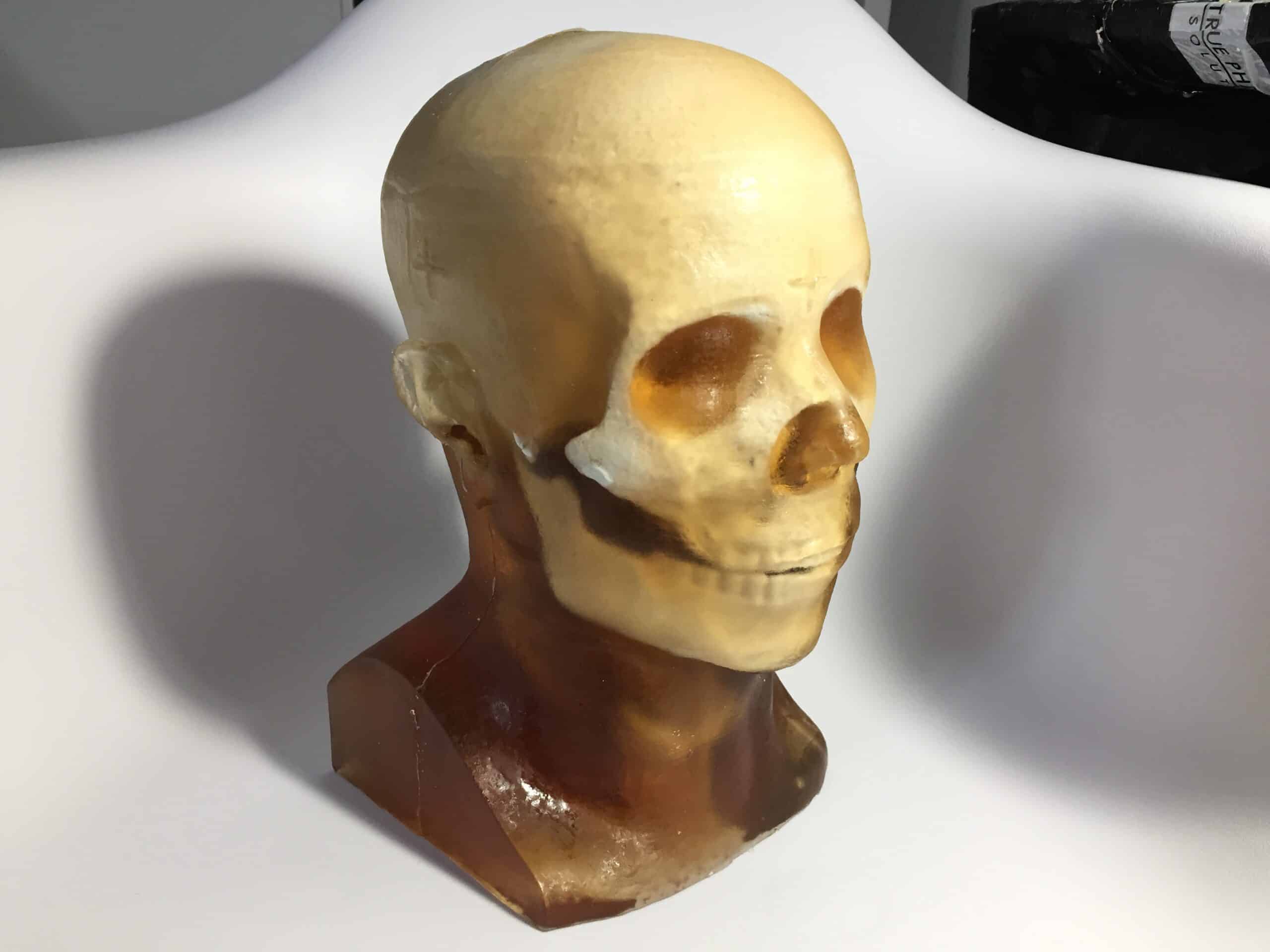
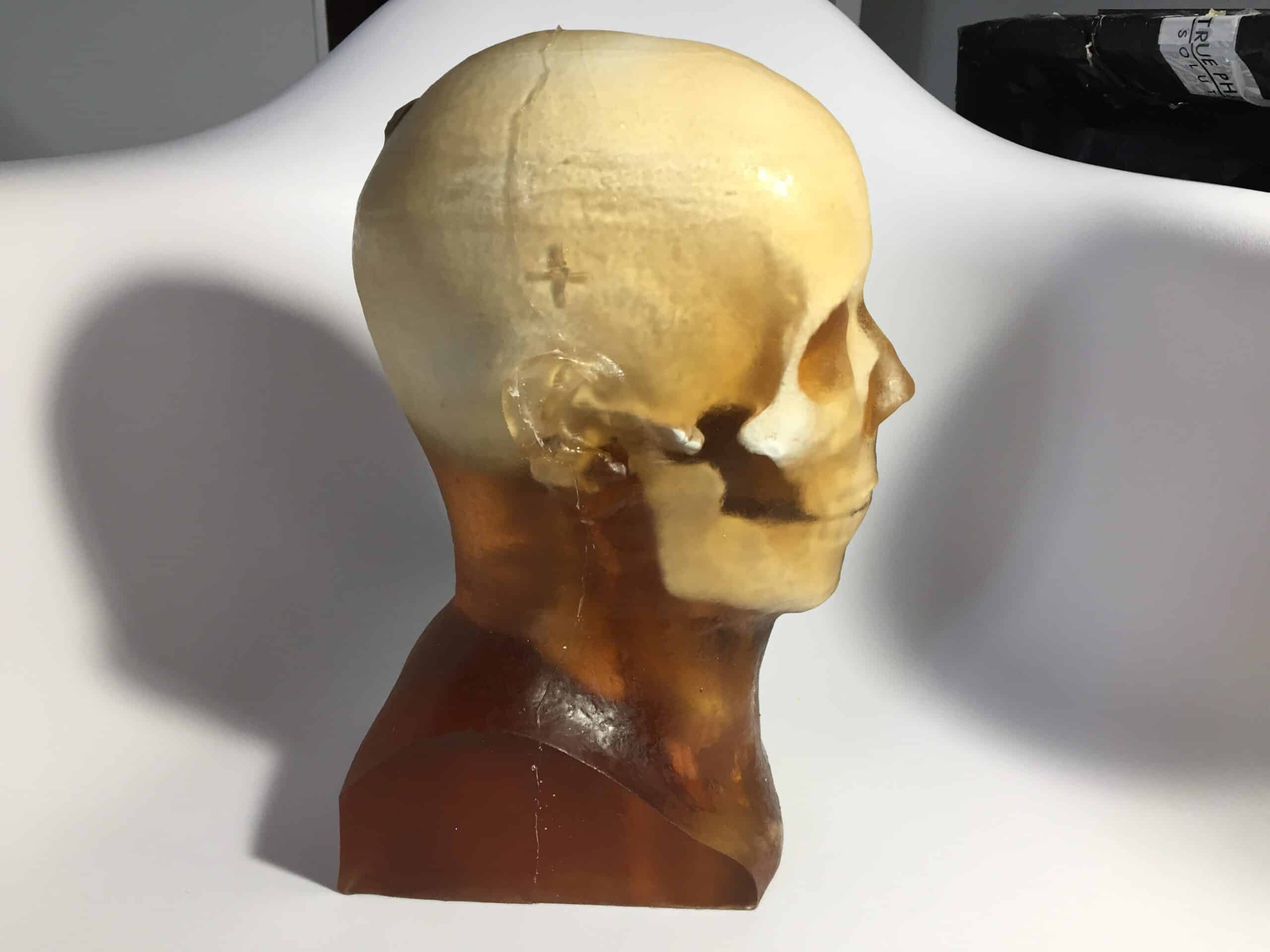
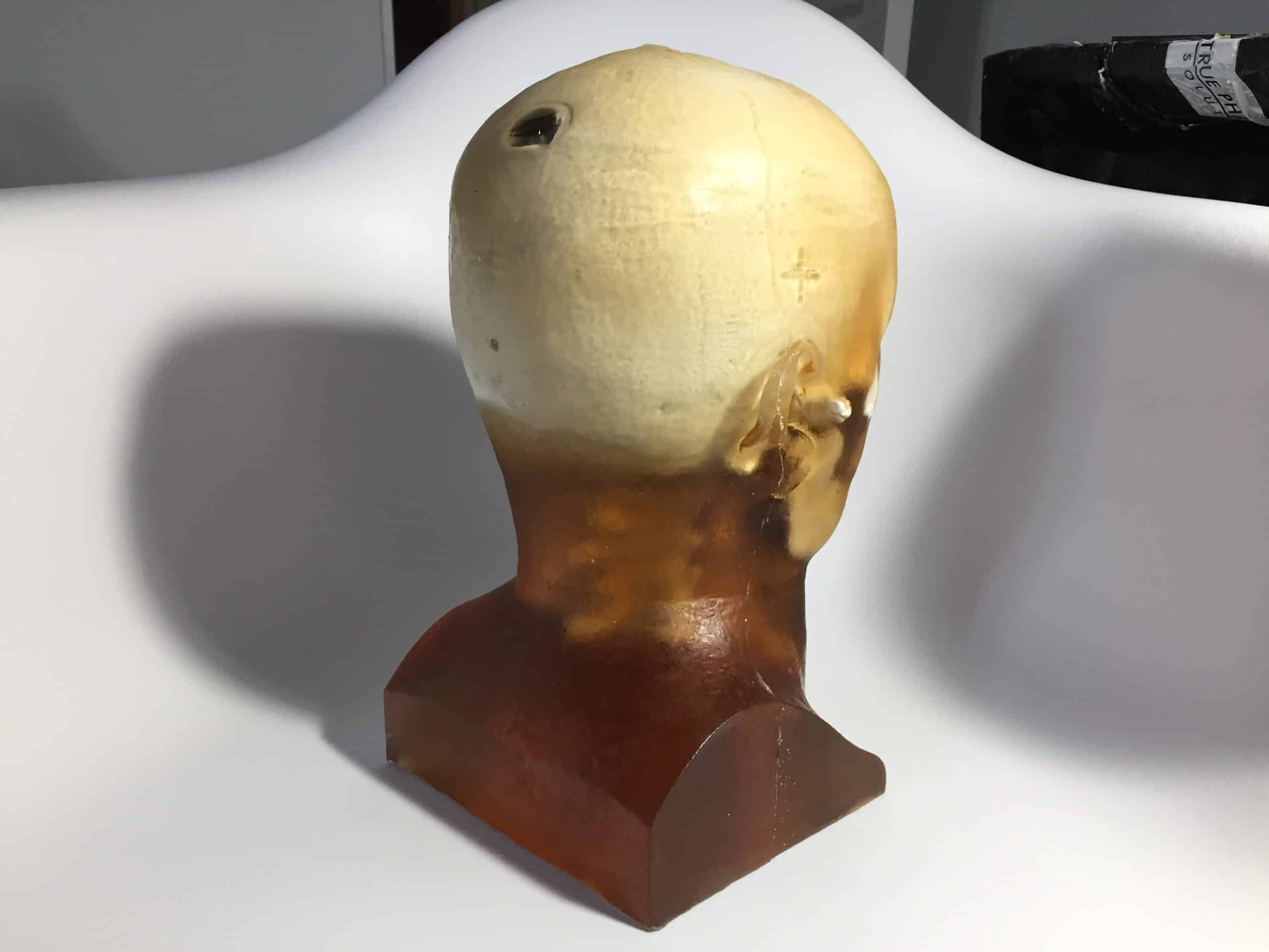
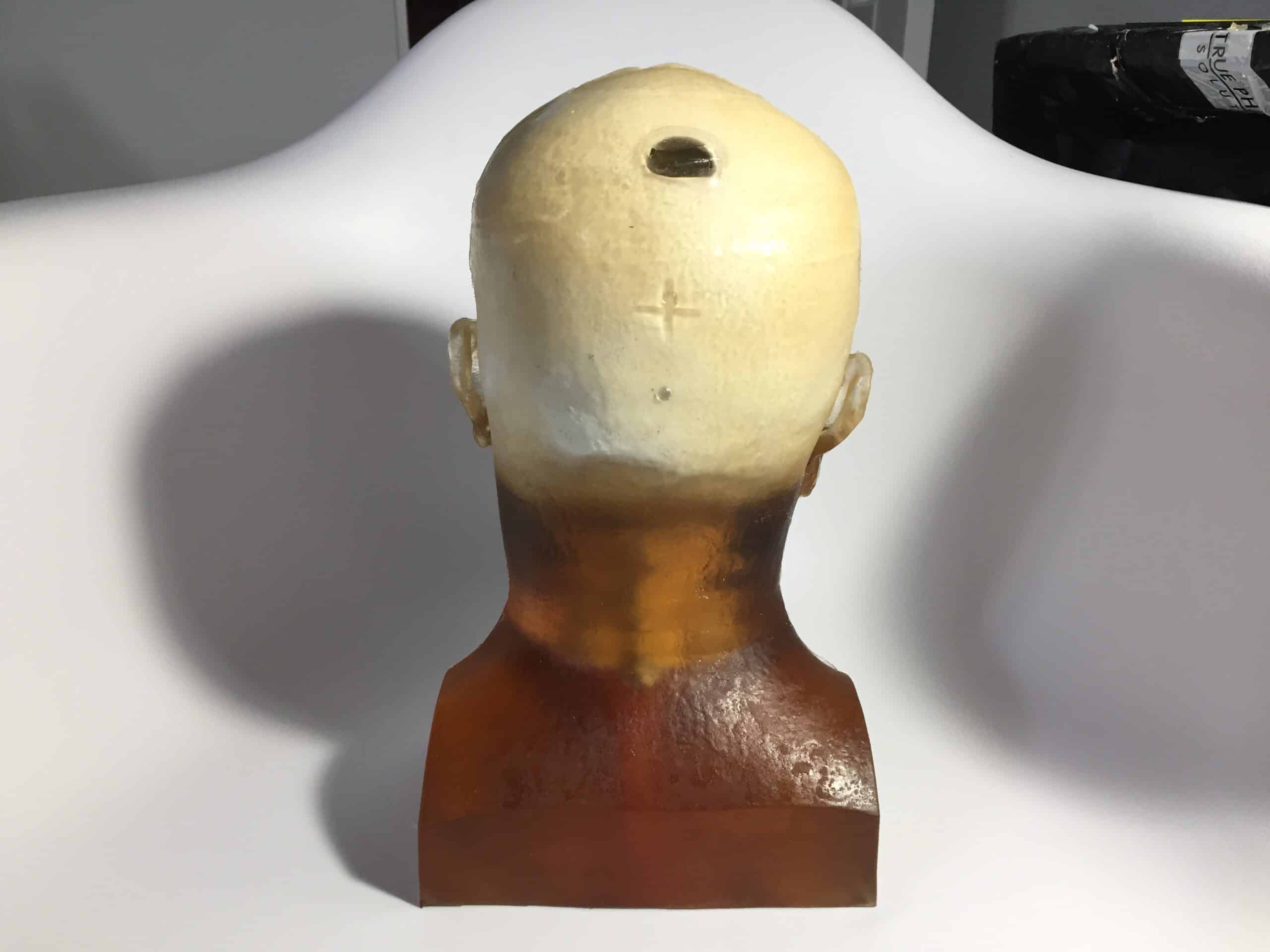
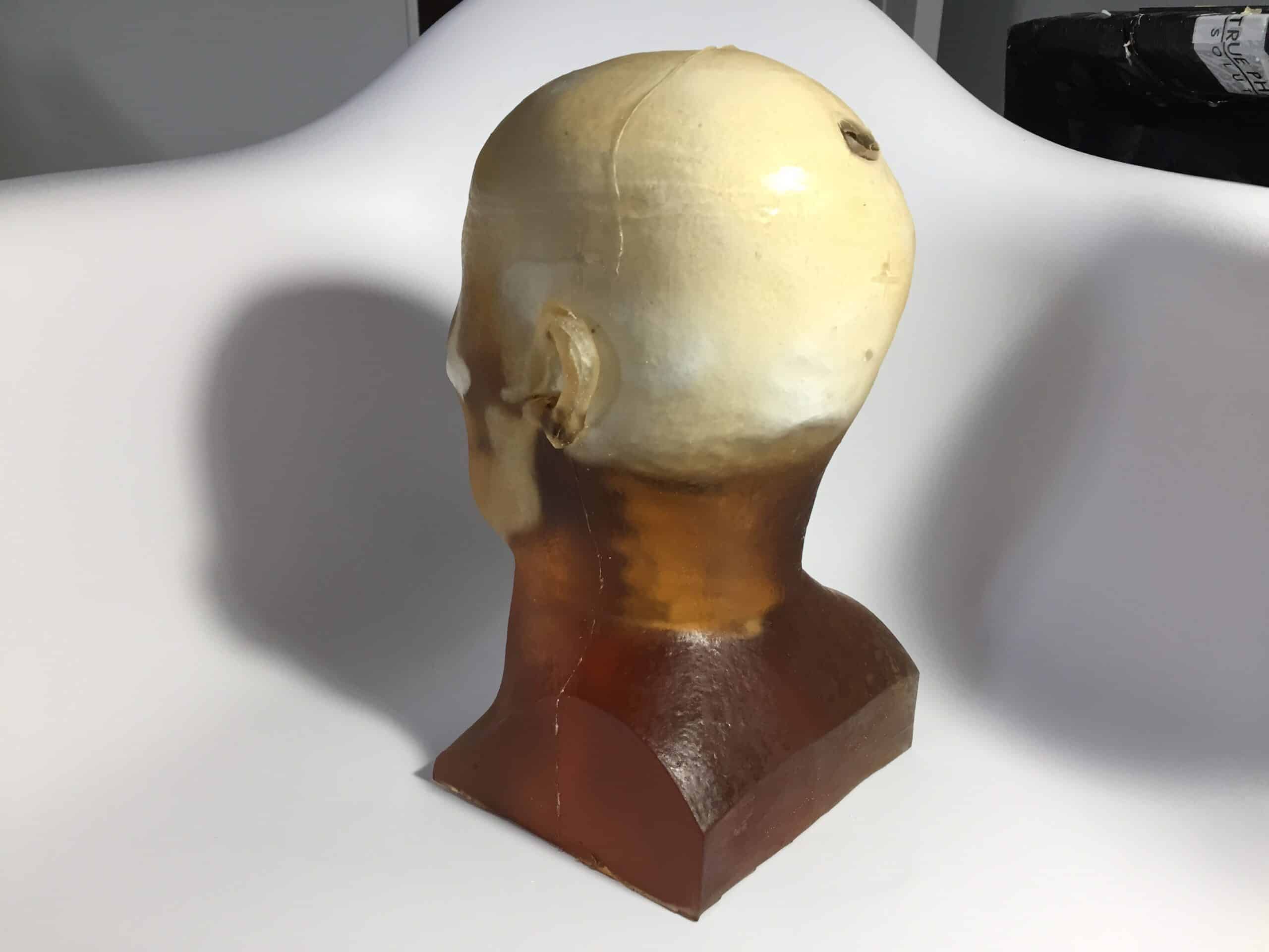
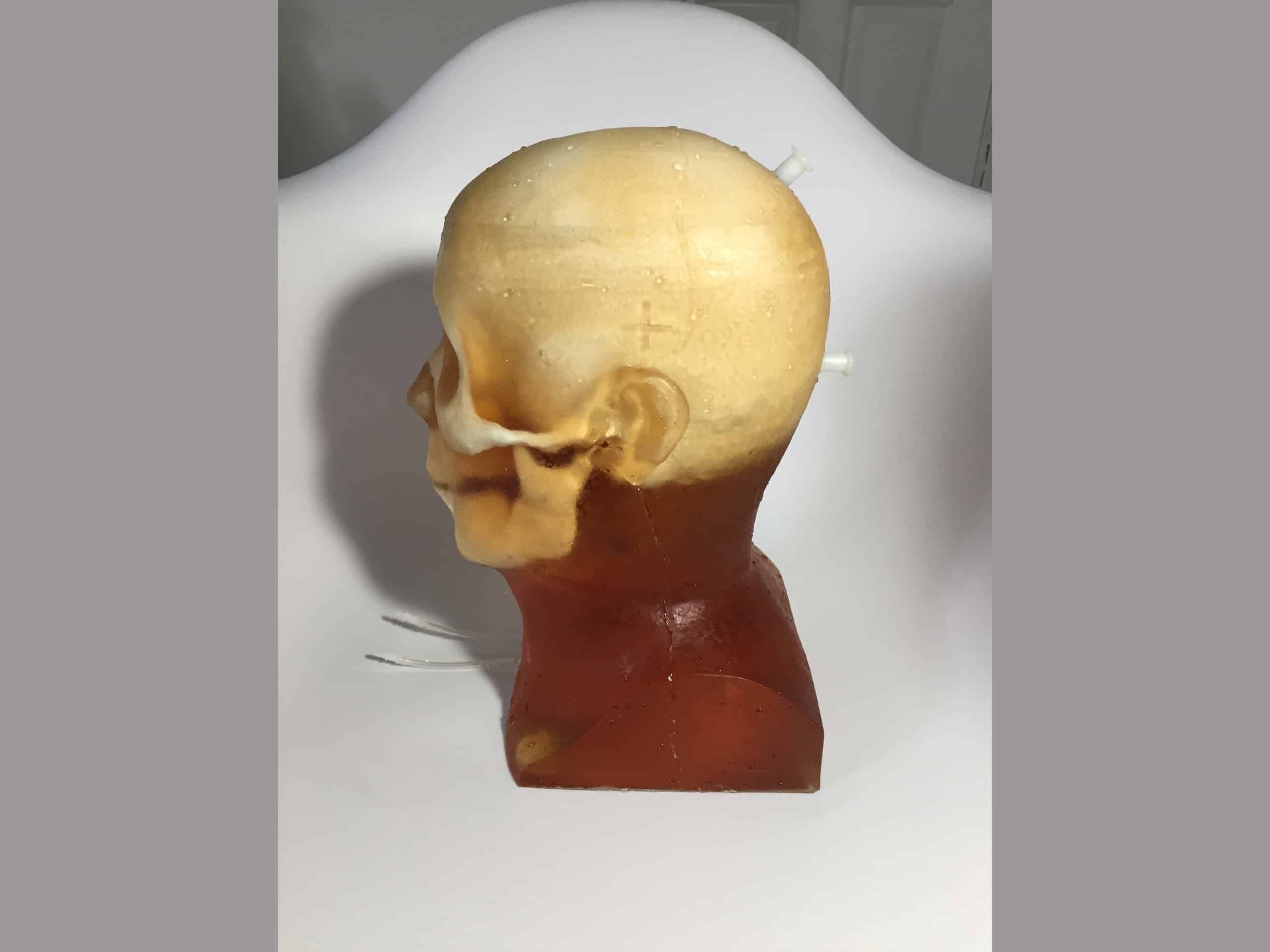
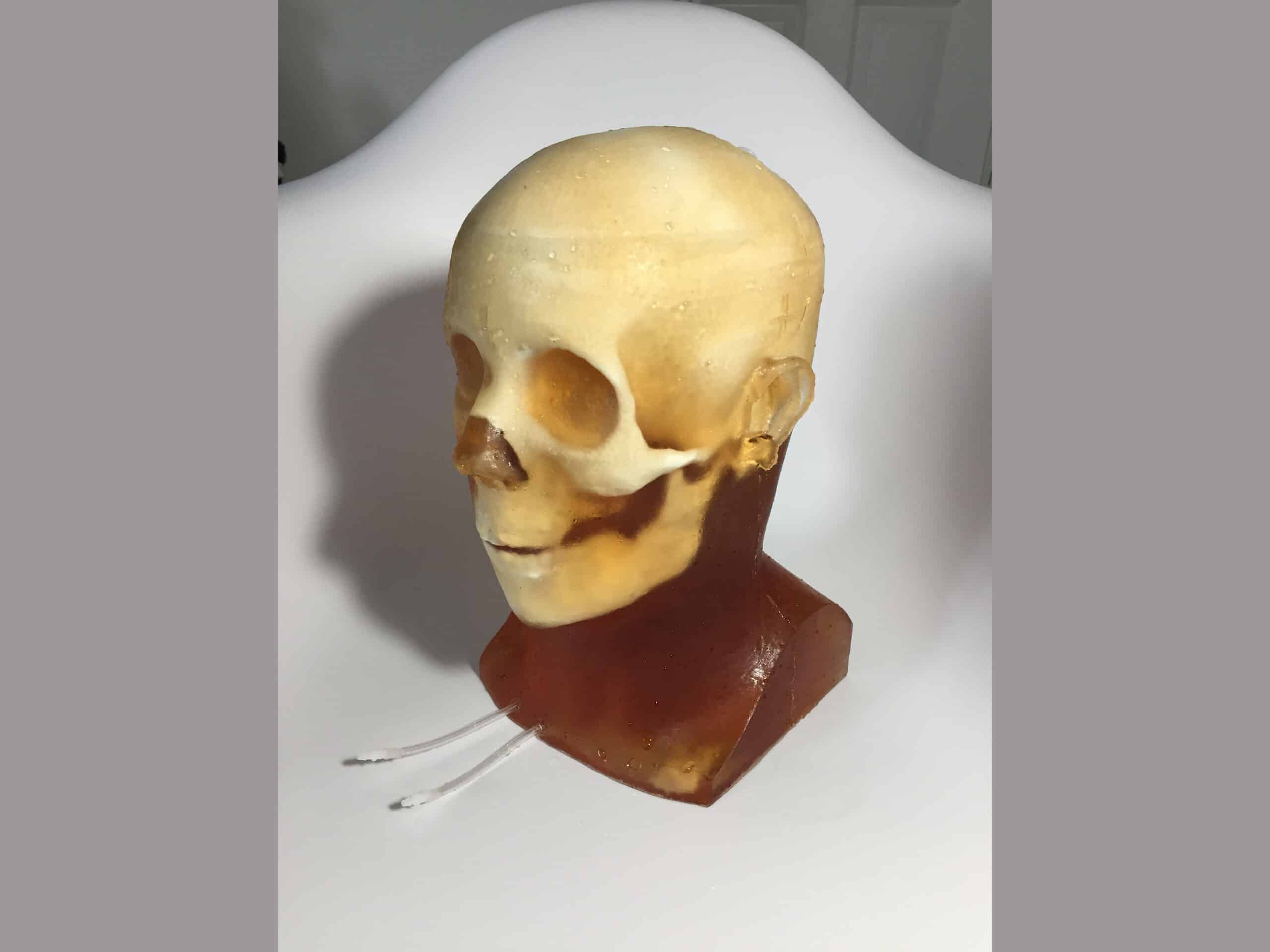
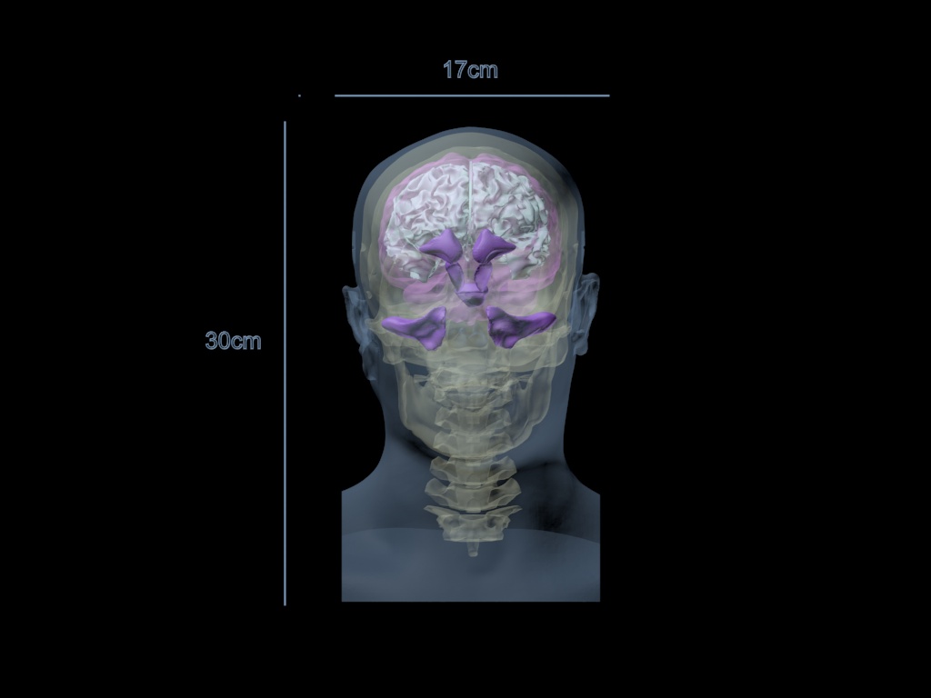
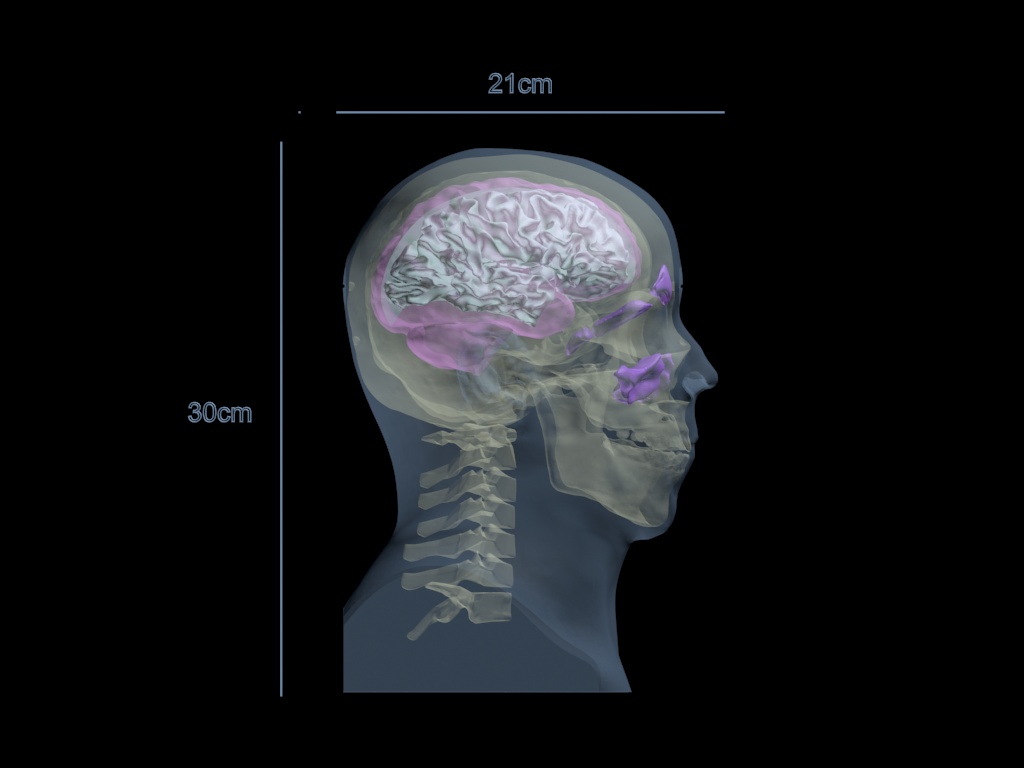
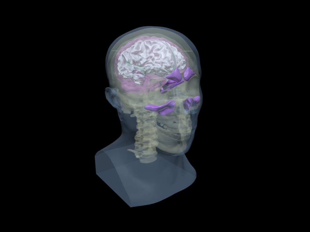
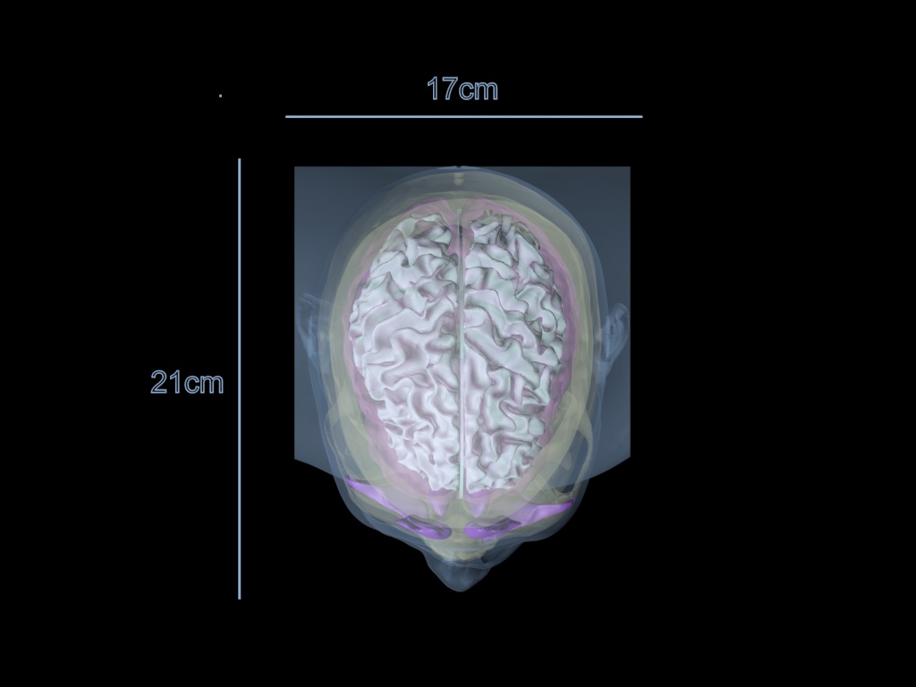
The Adult Head (Training, V1) is a radiology training phantom compatible with X-Ray/CT, MRI, and Ultrasound. The phantom’s skull bone has a realistic three-layered structure, including an inner diploe layer. The phantom also includes a brain with synthetic tissue and ventricles.
Head Models: Dynamic
Appearance: Amber Translucent
This product can be customized based on your requirements — contact us.
The Adult Head (Training, V1) is an X-ray/CT, MRI, and ultrasound-compatible training phantom used by radiology schools, medical schools & teaching hospitals for training their students & medical professionals in various scanning techniques. The skull bone has a realistic three-layered structure with an inner diploe layer. Thisphantom can be used with Barium-sulfate, Iodine-based, Gadolinium-based, Microbubble,and Iron Oxide-based contrast mediums.
In terms of MRI applications, the phantom tissues have realistic T2 relaxation time values, which makes this product the best fit for any T2-weighted MRI imaging methods. Very good results can also be achieved with proton-density imaging methods. The phantom can still be imaged with T1-weighted methods, but the T1 values are less realistic, and they are within the range of about 100 ms.
Note: By default, the phantom comes in a translucent amber appearance. The skin feature can be included upon request.
| Features | Dynamic, Adv. Head | Static Head | Dynamic Head | Standard Head | Training, V1 Head | Hematoma Head | Openable Head |
|---|---|---|---|---|---|---|---|
| Skull | ✓ | ✓ | ✓ | ✓ | ✓ | ✓ | ✓ |
| Openable and Removable Calvaria | ✓ | ✖ | ✖ | ✖ | ✖ | ✖ | ✓ |
| Cervical Spine | ✖ | ✓ | ✖ | ✖ | ✓ | ✓ | ✖ |
| Skin Mimicking Material | ✓ | ✓ | ✓ | ✓ | ✓ | ✓ | ✓ |
| Brain (Gray Matter) | ✓ | ✓ | ✓ | ✓ | ✓ | ✓ | ✖ |
| Brain Ventricles | ✓ | ✖ | ✖ | ✓ | ✖ | ✓ | ✖ |
| Brain Tumor | ✖ | ✖ | ✖ | ✓ | ✖ | ✖ | ✖ |
| Falx Cerebri | ✖ | ✖ | ✖ | ✓ | ✓ | ✓ | ✖ |
| White Matter | ✖ | ✖ | ✖ | ✖ | ✓ | ✖ | ✖ |
| Brain Vessels | ✓ ( {2mm, 3mm, 4mm} incorporated Circle of Willis ) | ✖ | ✓ ( {2mm, 3mm, 4mm} incorporated Circle of Willis ) | ✓ | ✖ | ✖ | ✖ |
| One Bifurcation | ✓ | ✖ | ✓ | ✓ | ✖ | ✖ | ✖ |
| One Stenosis | ✓ | ✖ | ✓ | ✖ | ✖ | ✖ | ✖ |
| Two Aneurysms | ✓ | ✖ | ✓ | ✖ | ✖ | ✖ | ✖ |
| Vessels Connected with the Neck | ✓ | ✖ | ✓ | ✓ | ✖ | ✖ | ✖ |
| Plastic Connectors | ✓ | ✖ | ✓ | ✖ | ✖ | ✖ | ✖ |
| Small Static Objects | ✖ | ✓ ( {0.5mm, 1mm, 2mm, 3mm Diameters} ) | ✖ | ✖ | ✖ | ✖ | ✖ |
| Realistic Air Cavities | ✖ | ✖ | ✖ | ✖ | ✓ | ✖ | ✖ |
| Alignment Markings | ✓ | ✓ | ✓ | ✓ | ✓ | ✓ | ✓ |
| Frontal Sinus with Air Gaps | ✖ | ✖ | ✖ | ✖ | ✓ | ✖ | ✖ |
| Hematoma | ✖ | ✖ | ✖ | ✖ | ✖ | ✓ | ✖ |
| Features<\/th>\n | Dynamic,\nAdv. Head<\/th>\n | Static\nHead<\/th>\n | Dynamic\nHead<\/th>\n | Standard\u00a0 \u00a0 \u00a0Head<\/th>\n | Training,\nV1 Head<\/th>\n | Hematoma Head<\/th>\n | Openable Head<\/th>\n<\/tr>\n<\/thead>\n | ||||||||||||||||||||||||||||||||||||||||||||||||||||||||||||||||||||||||||||||||||||||||||||||||||||||||||||||||||||||||||||||||||||||||||||||||||||||||||||||||||||||||||||||||||||||||||||||||||||||||||||||||||||||||||||||||||||||||||
|---|---|---|---|---|---|---|---|---|---|---|---|---|---|---|---|---|---|---|---|---|---|---|---|---|---|---|---|---|---|---|---|---|---|---|---|---|---|---|---|---|---|---|---|---|---|---|---|---|---|---|---|---|---|---|---|---|---|---|---|---|---|---|---|---|---|---|---|---|---|---|---|---|---|---|---|---|---|---|---|---|---|---|---|---|---|---|---|---|---|---|---|---|---|---|---|---|---|---|---|---|---|---|---|---|---|---|---|---|---|---|---|---|---|---|---|---|---|---|---|---|---|---|---|---|---|---|---|---|---|---|---|---|---|---|---|---|---|---|---|---|---|---|---|---|---|---|---|---|---|---|---|---|---|---|---|---|---|---|---|---|---|---|---|---|---|---|---|---|---|---|---|---|---|---|---|---|---|---|---|---|---|---|---|---|---|---|---|---|---|---|---|---|---|---|---|---|---|---|---|---|---|---|---|---|---|---|---|---|---|---|---|---|---|---|---|---|---|---|---|---|---|---|---|---|---|---|---|---|---|---|---|---|---|---|---|---|---|---|---|---|---|
| Skull<\/td>\n | \u2713<\/strong><\/td>\n| \u2713<\/span><\/td>\n | \u2713<\/span><\/td>\n | \u2713<\/span><\/td>\n | \u2713<\/span><\/td>\n | \u2713<\/span><\/td>\n | \u2713<\/span><\/td>\n<\/tr>\n | Openable and Removable Calvaria<\/td>\n | \u2713<\/strong><\/td>\n | \u2716<\/td>\n | \u2716<\/td>\n | \u2716<\/td>\n | \u2716<\/td>\n | \u2716<\/td>\n | \u2713<\/span><\/td>\n<\/tr>\n | Cervical Spine<\/td>\n | \u2716<\/span><\/td>\n | \u2713<\/span><\/td>\n | \u2716<\/span><\/td>\n | \u2716<\/span><\/td>\n | \u2713<\/span><\/td>\n | \u00a0\u2713<\/strong><\/td>\n | \u2716<\/span><\/td>\n<\/tr>\n | Skin Mimicking Material<\/td>\n | \u2713<\/strong><\/td>\n | \u2713<\/span><\/td>\n | \u2713<\/span><\/td>\n | \u2713<\/span><\/td>\n | \u2713<\/span><\/td>\n | \u2713<\/span><\/td>\n | \u2713<\/span><\/td>\n<\/tr>\n | Brain\n(Gray Matter)<\/td>\n | \u2713<\/strong><\/td>\n | \u2713<\/span><\/td>\n | \u2713<\/strong><\/td>\n | \u2713<\/span><\/td>\n | \u2713<\/span><\/td>\n | \u2713<\/span><\/td>\n | \u2716<\/span><\/td>\n<\/tr>\n | Brain Ventricles<\/td>\n | \u2713<\/strong><\/td>\n | \u2716<\/span><\/td>\n | \u2716<\/span><\/td>\n | \u2713<\/span><\/td>\n | \u2716<\/span><\/td>\n | \u2713<\/span><\/td>\n | \u2716<\/td>\n<\/tr>\n | Brain Tumor<\/td>\n | \u2716<\/span><\/td>\n | \u2716<\/span><\/td>\n | \u2716<\/span><\/td>\n | \u2713<\/span><\/td>\n | \u2716<\/span><\/td>\n | \u2716<\/span><\/td>\n | \u2716<\/span><\/td>\n<\/tr>\n | Falx Cerebri<\/td>\n | \u2716<\/span><\/td>\n | \u2716<\/span><\/td>\n | \u2716<\/span><\/td>\n | \u2713<\/span><\/td>\n | \u2713<\/span><\/td>\n | \u2713<\/span><\/td>\n | \u2716<\/span><\/td>\n<\/tr>\n | White Matter<\/td>\n | \u2716<\/span><\/td>\n | \u2716<\/span><\/td>\n | \u2716<\/span><\/td>\n | \u2716<\/span><\/td>\n | \u2713<\/span><\/td>\n | \u2716<\/span><\/td>\n | \u2716<\/td>\n<\/tr>\n | Brain Vessels<\/td>\n | \u2713 (\u00a0<\/strong>{2mm, 3mm, 4mm} incorporated Circle of Willis\u00a0<\/span>)<\/strong>\n<\/strong><\/td>\n | \u2716<\/span><\/td>\n | \u2713\u00a0<\/strong>(\u00a0<\/span>{<\/span>2mm, 3mm, 4mm} incorporated Circle of Willis\u00a0<\/span>)<\/span>\n<\/strong><\/td>\n | \u2713<\/strong><\/td>\n | \u2716<\/span><\/td>\n | \u2716<\/td>\n | \u2716<\/td>\n<\/tr>\n | One Bifurcation<\/td>\n | \u2713<\/span><\/td>\n | \u2716<\/span><\/td>\n | \u2713<\/span><\/td>\n | \u2713<\/strong><\/td>\n | \u2716<\/span><\/td>\n | \u2716<\/span><\/td>\n | \u2716<\/td>\n<\/tr>\n | One Stenosis<\/td>\n | \u2713<\/span><\/td>\n | \u2716<\/span><\/td>\n | \u2713<\/span><\/td>\n | \u2716<\/span><\/td>\n | \u2716<\/span><\/td>\n | \u2716<\/span><\/td>\n | \u2716<\/td>\n<\/tr>\n | Two Aneurysms<\/td>\n | \u2713<\/strong><\/td>\n | \u2716<\/span><\/td>\n | \u2713<\/span><\/td>\n | \u2716<\/td>\n | \u2716<\/td>\n | \u2716<\/td>\n | \u2716<\/td>\n<\/tr>\n | Vessels Connected with the Neck<\/td>\n | \u2713<\/span><\/td>\n | \u2716<\/strong><\/td>\n | \u2713<\/span><\/td>\n | \u2713<\/span><\/td>\n | \u2716<\/span><\/td>\n | \u2716<\/td>\n | \u2716<\/td>\n<\/tr>\n | Plastic Connectors<\/td>\n | \u2713<\/span><\/td>\n | \u2716<\/td>\n | \u2713<\/span><\/td>\n | \u2716<\/td>\n | \u2716<\/td>\n | \u2716<\/td>\n | \u2716<\/td>\n<\/tr>\n | Small Static Objects<\/td>\n | \u2716<\/td>\n | \u2713 <\/span>(<\/strong> {0.5mm, 1mm, 2mm, 3mm Diameters} )<\/b><\/span><\/td>\n | \u2716<\/td>\n | \u2716<\/span><\/td>\n | \u2716<\/td>\n | \u2716<\/td>\n | \u2716<\/td>\n<\/tr>\n | Realistic Air Cavities<\/td>\n | \u2716<\/td>\n | \u2716<\/td>\n | \u2716<\/td>\n | \u2716<\/td>\n | \u2713<\/strong><\/td>\n | \u2716<\/td>\n | \u2716<\/td>\n<\/tr>\n | Alignment Markings<\/td>\n | \u2713<\/span><\/td>\n | \u2713<\/span><\/td>\n | \u2713<\/span><\/td>\n | \u2713<\/span><\/td>\n | \u2713<\/strong><\/td>\n | \u2713<\/span><\/td>\n | \u2713<\/span><\/td>\n<\/tr>\n | Frontal Sinus with Air Gaps<\/td>\n | \u2716<\/td>\n | \u2716<\/td>\n | \u2716<\/td>\n | \u2716<\/td>\n | \u2713<\/strong><\/td>\n | \u2716<\/td>\n | \u2716<\/td>\n<\/tr>\n | Hematoma<\/td>\n | \u2716<\/td>\n | \u2716<\/td>\n | \u2716<\/td>\n | \u2716<\/td>\n | \u2716<\/td>\n | \u2713<\/strong><\/td>\n | \u2716<\/td>\n<\/tr>\n<\/tbody>\n<\/table>\n<\/div>\n"}}]}
| Scan Results:Coming Soon... In the meantime, please refer to our Radiographs page for imaging quality of our phantoms. Technical Properties:
Similar products
| ||||||||||||||||||||||||||||||||||||||||||||||||||||||||||||||||||||||||||||||||||