
X-Ray, CT, US, MRI Phantoms
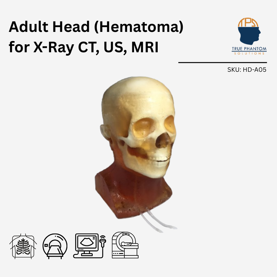
Adult Head (Hematoma) for X-Ray CT, US, MRI
$9,239
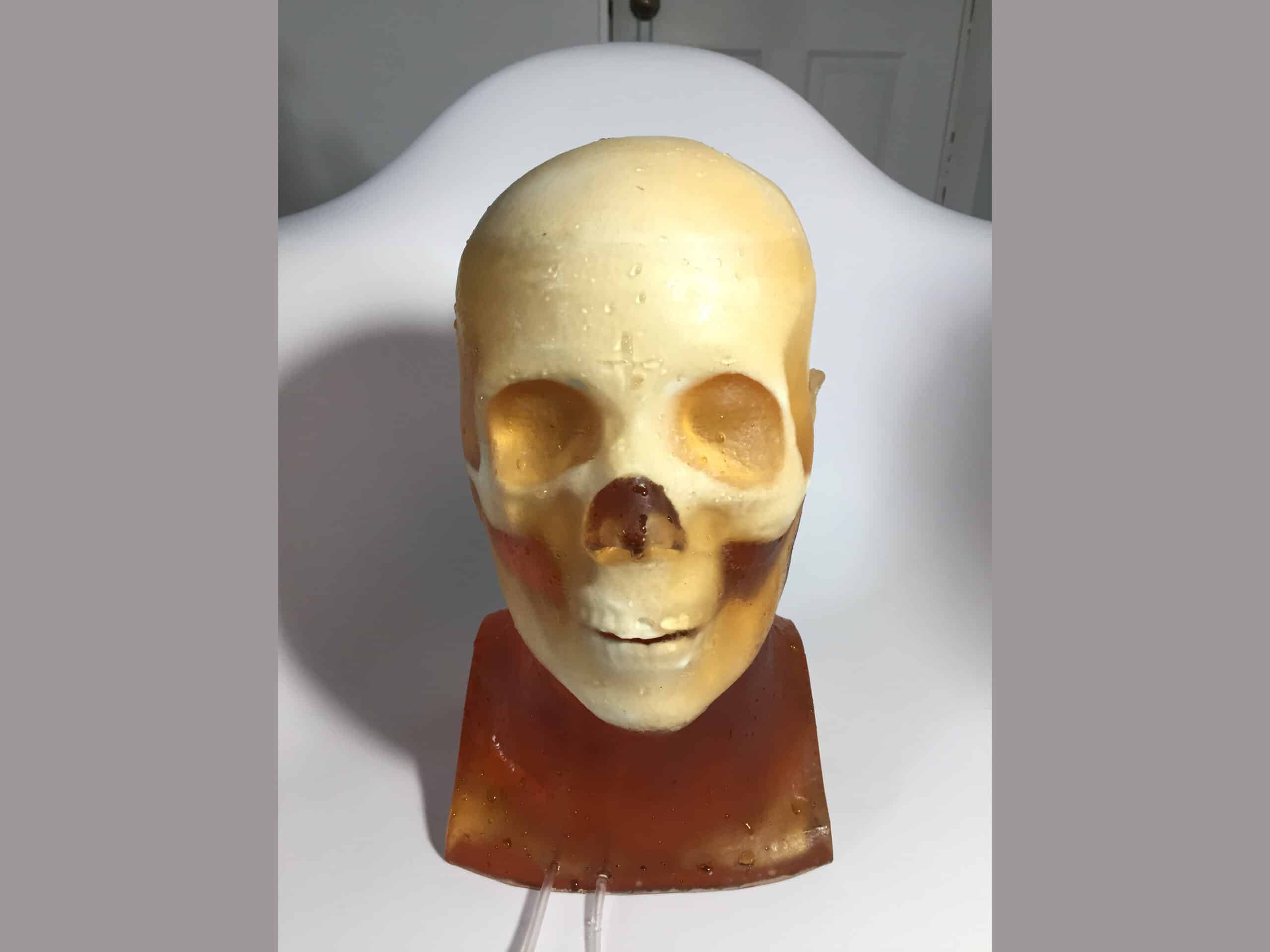
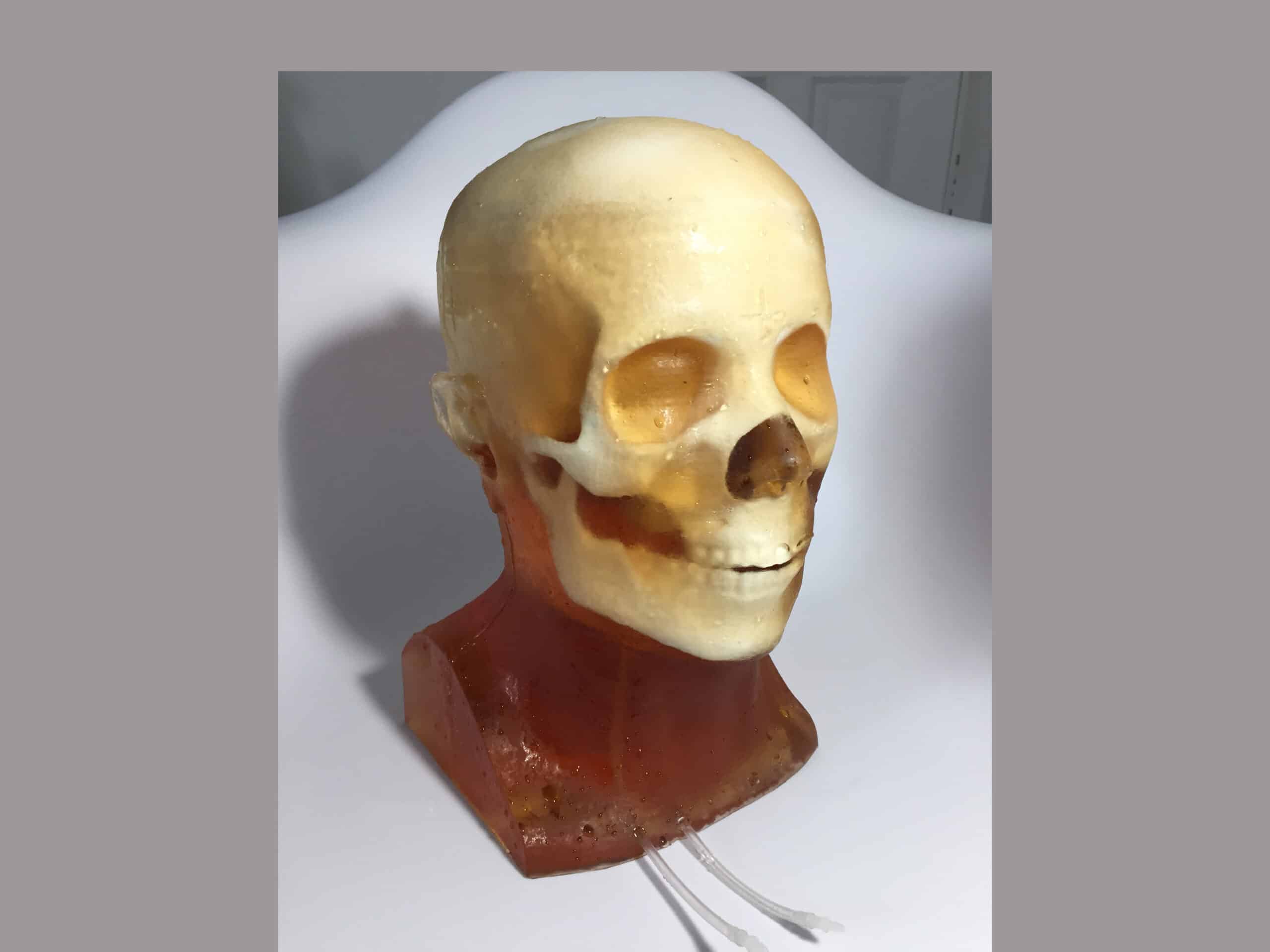
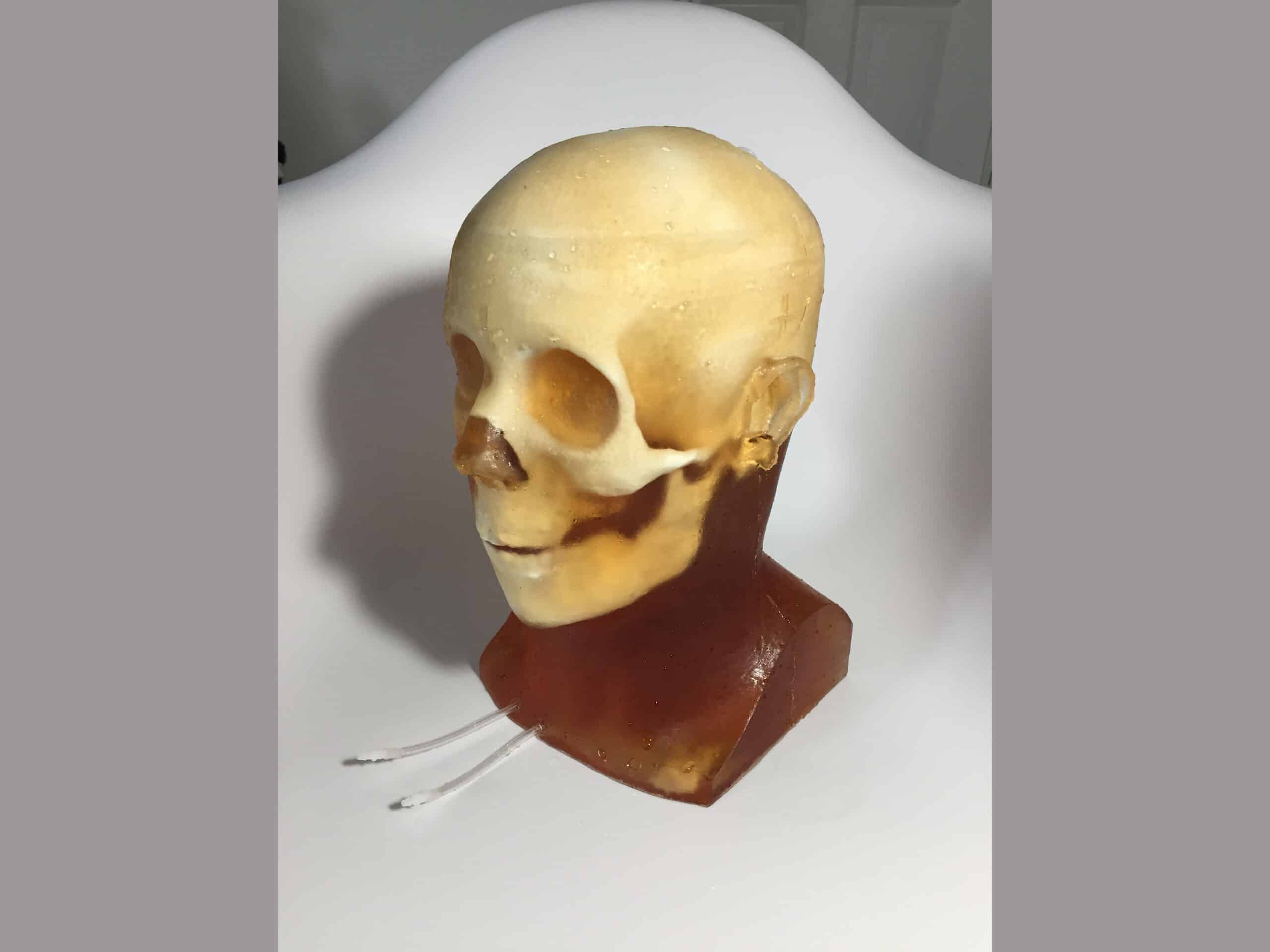
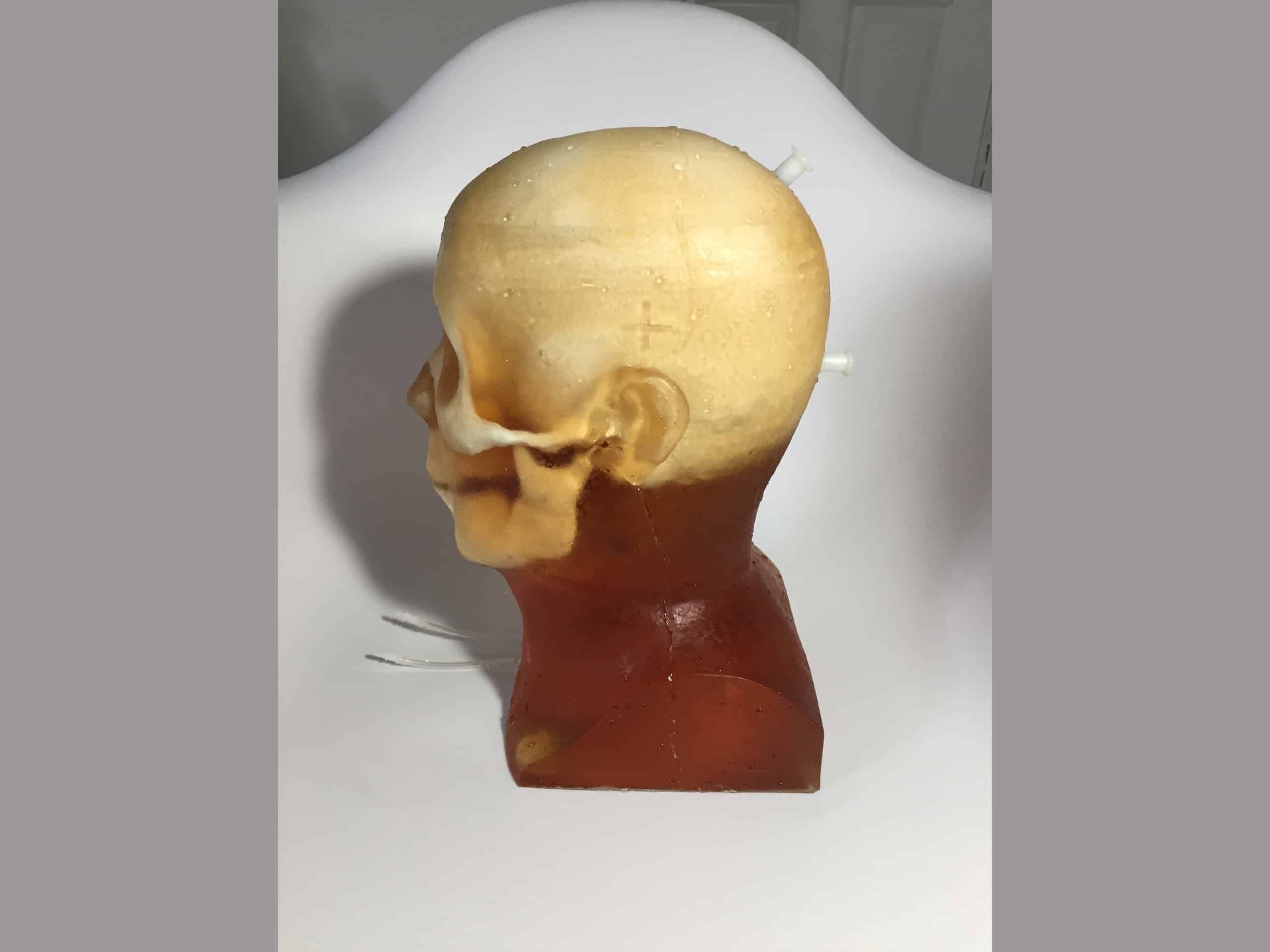
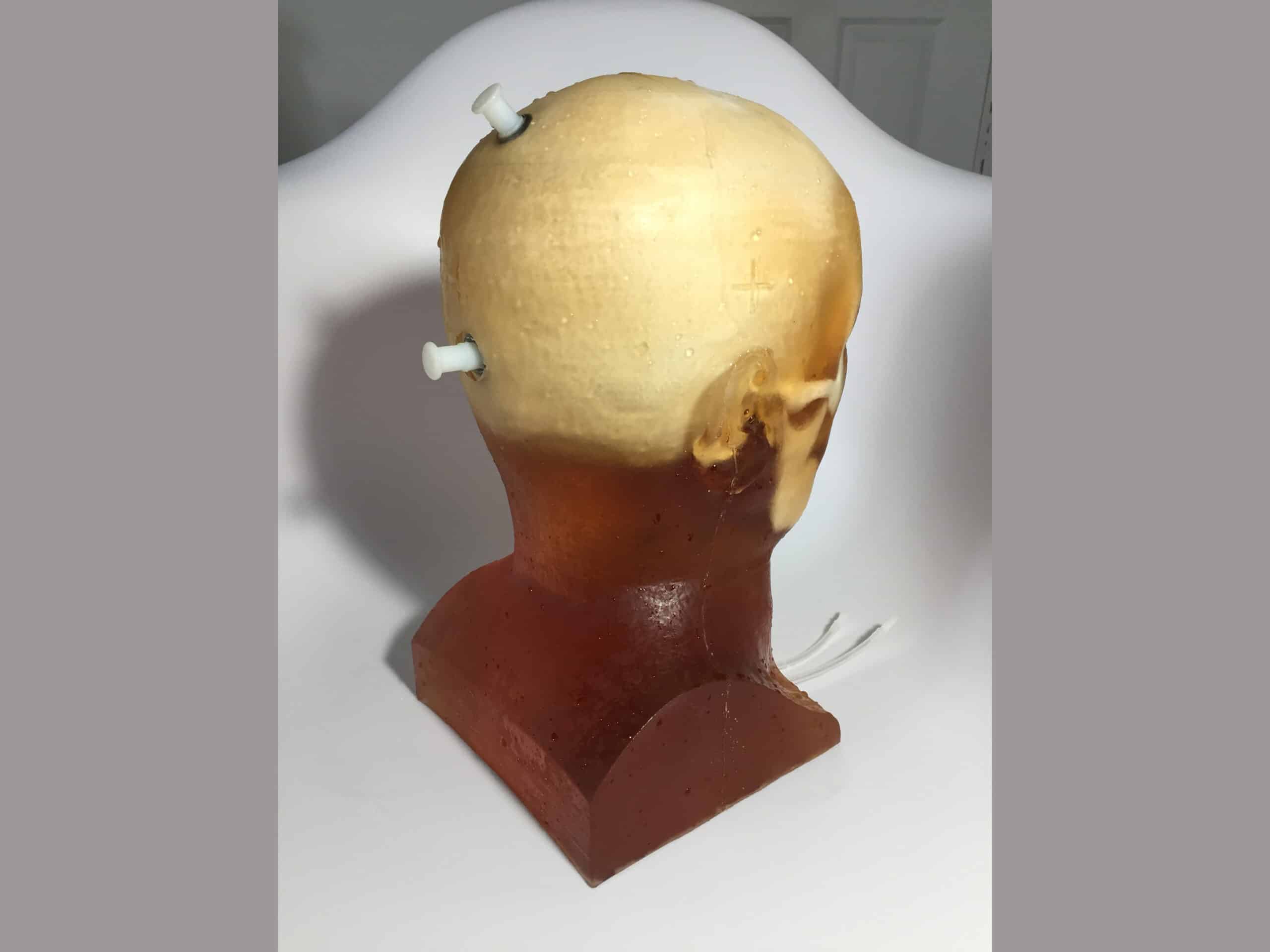
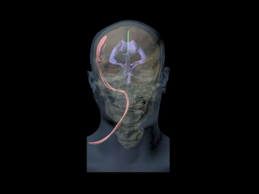
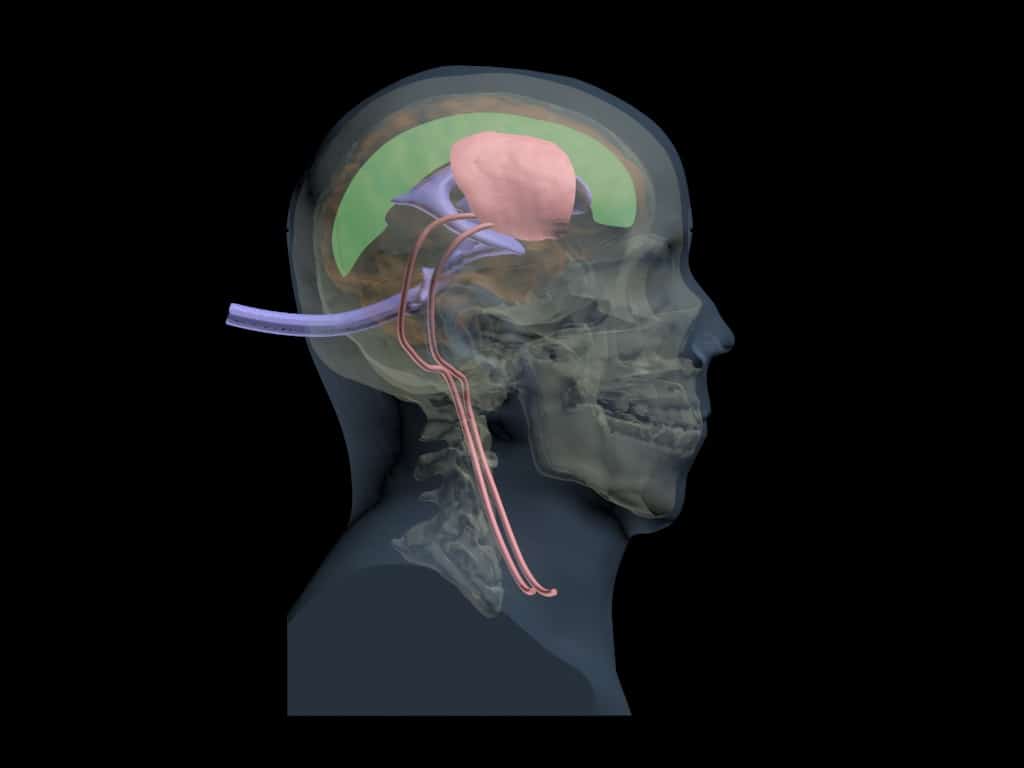
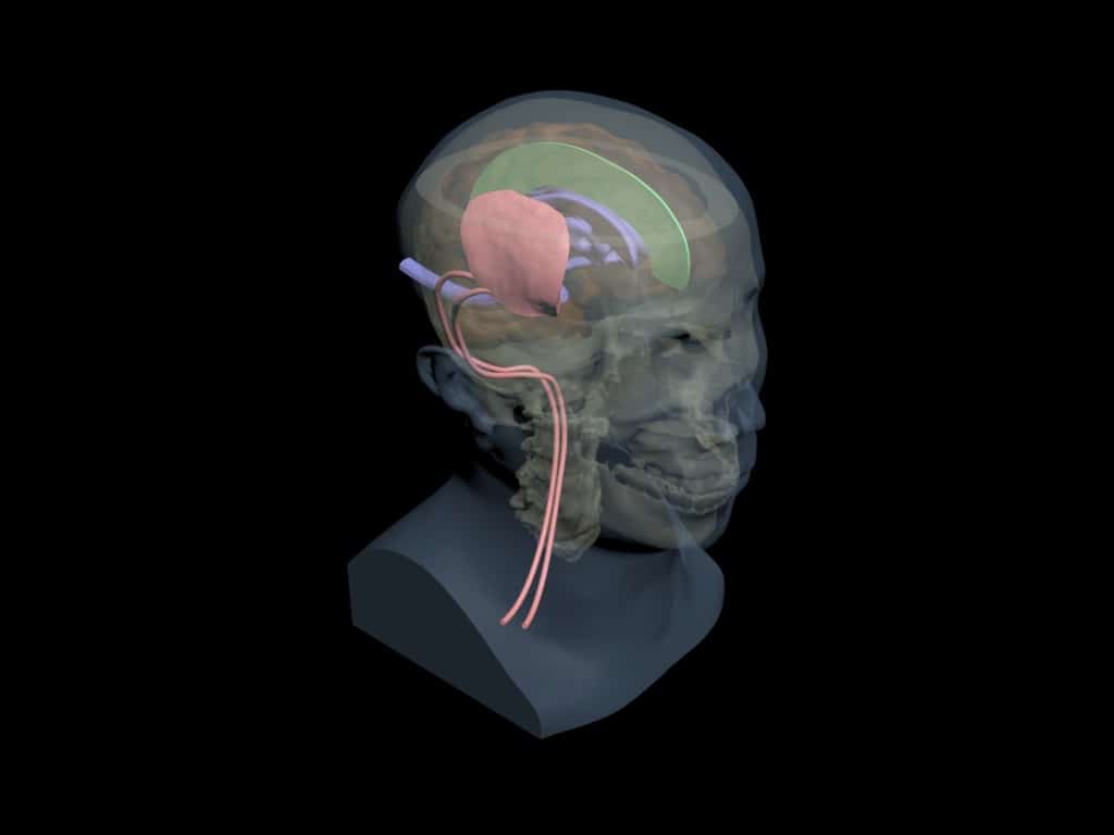
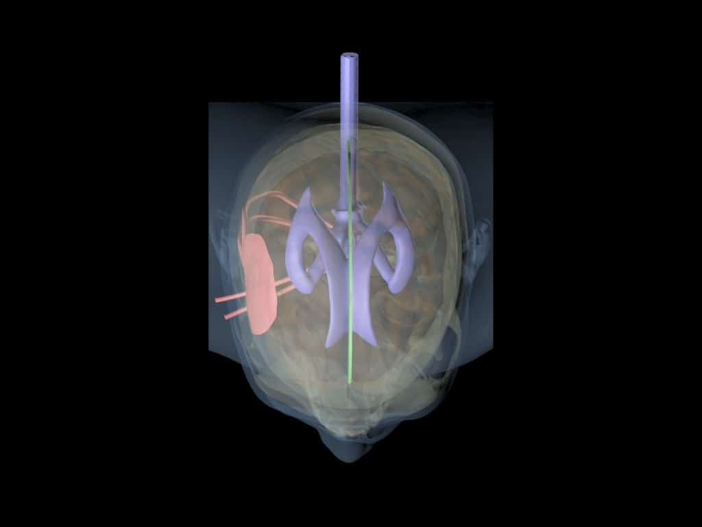
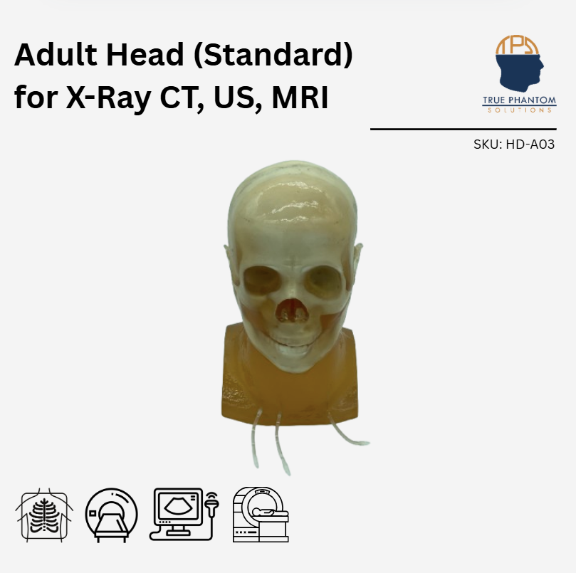
Our Adult Head (Hematoma) phantom is a cutting-edge medical training solution that seamlessly integrates with X-Ray CT, US, and MRI. This advanced phantom replicates the intricate anatomy of the human head, providing healthcare professionals with a realistic and immersive tool for skill development. This phantom is an excellent tool for applications relating to head trauma and hematomas. Upon special request, we can customize this head to mimic stroke, aneurysm, swelling and other such pathologies.
Head Models: Dynamic
Appearance: Amber Translucent
This product can be customized based on your requirements — contact us.
The Adult Head (Hematoma) phantom represents a revolutionary leap in medical training, offering a sophisticated solution that harmoniously interfaces with X-Ray CT, Ultrasound, and magnetic resonance imaging. Designed with precision, this phantom replicates the anatomy of the human head. This phantom can be used with Barium-sulfate, Iodine-based, Gadolinium-based, Microbubble, and Iron Oxide-based contrast mediums.
In terms of MRI applications, the phantom tissues have realistic T2 relaxation time values, which makes this product to be the best fit for any T2-weighted MRI imaging methods. Very good results can also be achieved with Proton Density imaging methods. The phantom can still be imaged with T1-weighted methods, but the T1 values are less realistic, and they are within the range of about 100 ms.
This advanced phantom serves as an exceptional training instrument for a range of medical applications, particularly those related to head trauma, hematomas, stroke, and aneurysms. By providing a lifelike simulation of various clinical scenarios, it empowers practitioners to refine their expertise in a risk-free environment.
| Features | Dynamic, Adv. Head | Static Head | Dynamic Head | Standard Head | Training, V1 Head | Hematoma Head | Openable Head |
|---|---|---|---|---|---|---|---|
| Skull | ✓ | ✓ | ✓ | ✓ | ✓ | ✓ | ✓ |
| Openable and Removable Calvaria | ✓ | ✖ | ✖ | ✖ | ✖ | ✖ | ✓ |
| Cervical Spine | ✖ | ✓ | ✖ | ✖ | ✓ | ✓ | ✖ |
| Skin Mimicking Material | ✓ | ✓ | ✓ | ✓ | ✓ | ✓ | ✓ |
| Brain (Gray Matter) | ✓ | ✓ | ✓ | ✓ | ✓ | ✓ | ✖ |
| Brain Ventricles | ✓ | ✖ | ✖ | ✓ | ✖ | ✓ | ✖ |
| Brain Tumor | ✖ | ✖ | ✖ | ✓ | ✖ | ✖ | ✖ |
| Falx Cerebri | ✖ | ✖ | ✖ | ✓ | ✓ | ✓ | ✖ |
| White Matter | ✖ | ✖ | ✖ | ✖ | ✓ | ✖ | ✖ |
| Brain Vessels | ✓ ( {2mm, 3mm, 4mm} incorporated Circle of Willis ) | ✖ | ✓ ( {2mm, 3mm, 4mm} incorporated Circle of Willis ) | ✓ | ✖ | ✖ | ✖ |
| One Bifurcation | ✓ | ✖ | ✓ | ✓ | ✖ | ✖ | ✖ |
| One Stenosis | ✓ | ✖ | ✓ | ✖ | ✖ | ✖ | ✖ |
| Two Aneurysms | ✓ | ✖ | ✓ | ✖ | ✖ | ✖ | ✖ |
| Vessels Connected with the Neck | ✓ | ✖ | ✓ | ✓ | ✖ | ✖ | ✖ |
| Plastic Connectors | ✓ | ✖ | ✓ | ✖ | ✖ | ✖ | ✖ |
| Small Static Objects | ✖ | ✓ ( {0.5mm, 1mm, 2mm, 3mm Diameters} ) | ✖ | ✖ | ✖ | ✖ | ✖ |
| Realistic Air Cavities | ✖ | ✖ | ✖ | ✖ | ✓ | ✖ | ✖ |
| Alignment Markings | ✓ | ✓ | ✓ | ✓ | ✓ | ✓ | ✓ |
| Frontal Sinus with Air Gaps | ✖ | ✖ | ✖ | ✖ | ✓ | ✖ | ✖ |
| Hematoma | ✖ | ✖ | ✖ | ✖ | ✖ | ✓ | ✖ |
| Features<\/th>\n | Dynamic,\nAdv. Head<\/th>\n | Static\nHead<\/th>\n | Dynamic\nHead<\/th>\n | Standard\u00a0 \u00a0 \u00a0Head<\/th>\n | Training,\nV1 Head<\/th>\n | Hematoma Head<\/th>\n | Openable Head<\/th>\n<\/tr>\n<\/thead>\n | ||||||||||||||||||||||||||||||||||||||||||||||||||||||||||||||||||||||||||||||||||||||||||||||||||||||||||||||||||||||||||||||||||||||||||||||||||||||||||||||||||||||||||||||||||||||||||||||||||||||||||||||||||||||||||||||||||||||||||
|---|---|---|---|---|---|---|---|---|---|---|---|---|---|---|---|---|---|---|---|---|---|---|---|---|---|---|---|---|---|---|---|---|---|---|---|---|---|---|---|---|---|---|---|---|---|---|---|---|---|---|---|---|---|---|---|---|---|---|---|---|---|---|---|---|---|---|---|---|---|---|---|---|---|---|---|---|---|---|---|---|---|---|---|---|---|---|---|---|---|---|---|---|---|---|---|---|---|---|---|---|---|---|---|---|---|---|---|---|---|---|---|---|---|---|---|---|---|---|---|---|---|---|---|---|---|---|---|---|---|---|---|---|---|---|---|---|---|---|---|---|---|---|---|---|---|---|---|---|---|---|---|---|---|---|---|---|---|---|---|---|---|---|---|---|---|---|---|---|---|---|---|---|---|---|---|---|---|---|---|---|---|---|---|---|---|---|---|---|---|---|---|---|---|---|---|---|---|---|---|---|---|---|---|---|---|---|---|---|---|---|---|---|---|---|---|---|---|---|---|---|---|---|---|---|---|---|---|---|---|---|---|---|---|---|---|---|---|---|---|---|---|
| Skull<\/td>\n | \u2713<\/strong><\/td>\n| \u2713<\/span><\/td>\n | \u2713<\/span><\/td>\n | \u2713<\/span><\/td>\n | \u2713<\/span><\/td>\n | \u2713<\/span><\/td>\n | \u2713<\/span><\/td>\n<\/tr>\n | Openable and Removable Calvaria<\/td>\n | \u2713<\/strong><\/td>\n | \u2716<\/td>\n | \u2716<\/td>\n | \u2716<\/td>\n | \u2716<\/td>\n | \u2716<\/td>\n | \u2713<\/span><\/td>\n<\/tr>\n | Cervical Spine<\/td>\n | \u2716<\/span><\/td>\n | \u2713<\/span><\/td>\n | \u2716<\/span><\/td>\n | \u2716<\/span><\/td>\n | \u2713<\/span><\/td>\n | \u00a0\u2713<\/strong><\/td>\n | \u2716<\/span><\/td>\n<\/tr>\n | Skin Mimicking Material<\/td>\n | \u2713<\/strong><\/td>\n | \u2713<\/span><\/td>\n | \u2713<\/span><\/td>\n | \u2713<\/span><\/td>\n | \u2713<\/span><\/td>\n | \u2713<\/span><\/td>\n | \u2713<\/span><\/td>\n<\/tr>\n | Brain\n(Gray Matter)<\/td>\n | \u2713<\/strong><\/td>\n | \u2713<\/span><\/td>\n | \u2713<\/strong><\/td>\n | \u2713<\/span><\/td>\n | \u2713<\/span><\/td>\n | \u2713<\/span><\/td>\n | \u2716<\/span><\/td>\n<\/tr>\n | Brain Ventricles<\/td>\n | \u2713<\/strong><\/td>\n | \u2716<\/span><\/td>\n | \u2716<\/span><\/td>\n | \u2713<\/span><\/td>\n | \u2716<\/span><\/td>\n | \u2713<\/span><\/td>\n | \u2716<\/td>\n<\/tr>\n | Brain Tumor<\/td>\n | \u2716<\/span><\/td>\n | \u2716<\/span><\/td>\n | \u2716<\/span><\/td>\n | \u2713<\/span><\/td>\n | \u2716<\/span><\/td>\n | \u2716<\/span><\/td>\n | \u2716<\/span><\/td>\n<\/tr>\n | Falx Cerebri<\/td>\n | \u2716<\/span><\/td>\n | \u2716<\/span><\/td>\n | \u2716<\/span><\/td>\n | \u2713<\/span><\/td>\n | \u2713<\/span><\/td>\n | \u2713<\/span><\/td>\n | \u2716<\/span><\/td>\n<\/tr>\n | White Matter<\/td>\n | \u2716<\/span><\/td>\n | \u2716<\/span><\/td>\n | \u2716<\/span><\/td>\n | \u2716<\/span><\/td>\n | \u2713<\/span><\/td>\n | \u2716<\/span><\/td>\n | \u2716<\/td>\n<\/tr>\n | Brain Vessels<\/td>\n | \u2713 (\u00a0<\/strong>{2mm, 3mm, 4mm} incorporated Circle of Willis\u00a0<\/span>)<\/strong>\n<\/strong><\/td>\n | \u2716<\/span><\/td>\n | \u2713\u00a0<\/strong>(\u00a0<\/span>{<\/span>2mm, 3mm, 4mm} incorporated Circle of Willis\u00a0<\/span>)<\/span>\n<\/strong><\/td>\n | \u2713<\/strong><\/td>\n | \u2716<\/span><\/td>\n | \u2716<\/td>\n | \u2716<\/td>\n<\/tr>\n | One Bifurcation<\/td>\n | \u2713<\/span><\/td>\n | \u2716<\/span><\/td>\n | \u2713<\/span><\/td>\n | \u2713<\/strong><\/td>\n | \u2716<\/span><\/td>\n | \u2716<\/span><\/td>\n | \u2716<\/td>\n<\/tr>\n | One Stenosis<\/td>\n | \u2713<\/span><\/td>\n | \u2716<\/span><\/td>\n | \u2713<\/span><\/td>\n | \u2716<\/span><\/td>\n | \u2716<\/span><\/td>\n | \u2716<\/span><\/td>\n | \u2716<\/td>\n<\/tr>\n | Two Aneurysms<\/td>\n | \u2713<\/strong><\/td>\n | \u2716<\/span><\/td>\n | \u2713<\/span><\/td>\n | \u2716<\/td>\n | \u2716<\/td>\n | \u2716<\/td>\n | \u2716<\/td>\n<\/tr>\n | Vessels Connected with the Neck<\/td>\n | \u2713<\/span><\/td>\n | \u2716<\/strong><\/td>\n | \u2713<\/span><\/td>\n | \u2713<\/span><\/td>\n | \u2716<\/span><\/td>\n | \u2716<\/td>\n | \u2716<\/td>\n<\/tr>\n | Plastic Connectors<\/td>\n | \u2713<\/span><\/td>\n | \u2716<\/td>\n | \u2713<\/span><\/td>\n | \u2716<\/td>\n | \u2716<\/td>\n | \u2716<\/td>\n | \u2716<\/td>\n<\/tr>\n | Small Static Objects<\/td>\n | \u2716<\/td>\n | \u2713 <\/span>(<\/strong> {0.5mm, 1mm, 2mm, 3mm Diameters} )<\/b><\/span><\/td>\n | \u2716<\/td>\n | \u2716<\/span><\/td>\n | \u2716<\/td>\n | \u2716<\/td>\n | \u2716<\/td>\n<\/tr>\n | Realistic Air Cavities<\/td>\n | \u2716<\/td>\n | \u2716<\/td>\n | \u2716<\/td>\n | \u2716<\/td>\n | \u2713<\/strong><\/td>\n | \u2716<\/td>\n | \u2716<\/td>\n<\/tr>\n | Alignment Markings<\/td>\n | \u2713<\/span><\/td>\n | \u2713<\/span><\/td>\n | \u2713<\/span><\/td>\n | \u2713<\/span><\/td>\n | \u2713<\/strong><\/td>\n | \u2713<\/span><\/td>\n | \u2713<\/span><\/td>\n<\/tr>\n | Frontal Sinus with Air Gaps<\/td>\n | \u2716<\/td>\n | \u2716<\/td>\n | \u2716<\/td>\n | \u2716<\/td>\n | \u2713<\/strong><\/td>\n | \u2716<\/td>\n | \u2716<\/td>\n<\/tr>\n | Hematoma<\/td>\n | \u2716<\/td>\n | \u2716<\/td>\n | \u2716<\/td>\n | \u2716<\/td>\n | \u2716<\/td>\n | \u2713<\/strong><\/td>\n | \u2716<\/td>\n<\/tr>\n<\/tbody>\n<\/table>\n<\/div>"}}]}
| Scan Results:Coming Soon... For more imaging quality references, please see our Radiographs page. Technical Properties:
Similar products
| ||||||||||||||||||||||||||||||||||||||||||||||||||||||||||||||||||||||||||||||||||