




X-Ray, CT, US, MRI Phantoms
Head QC1 for X-Ray CT, US, MRI
$11,600
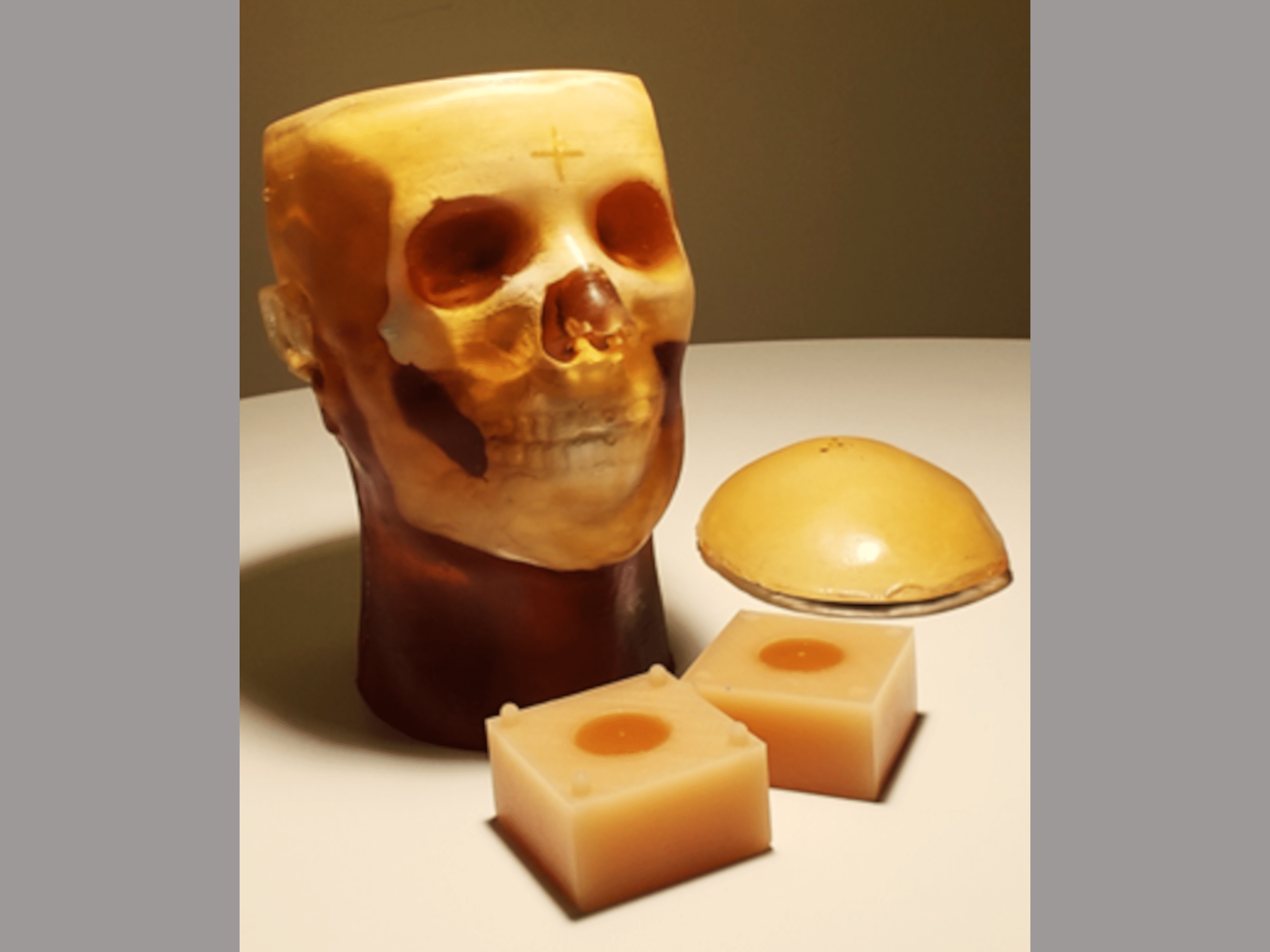
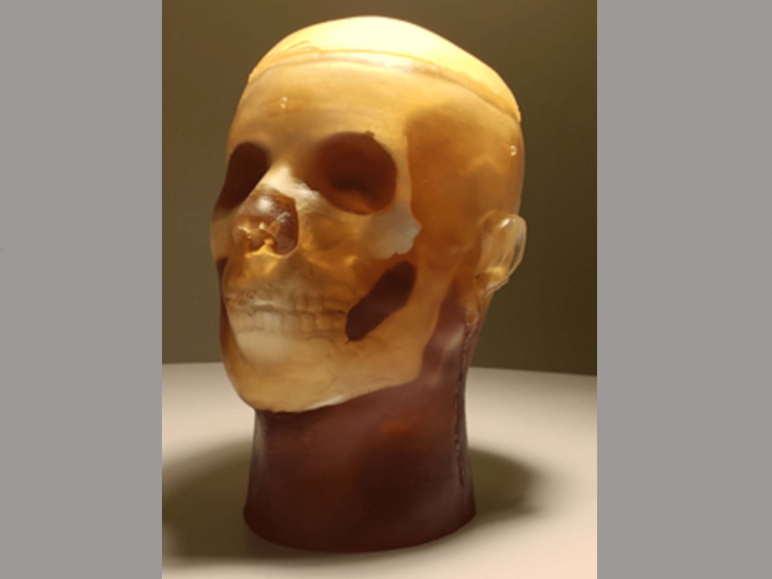
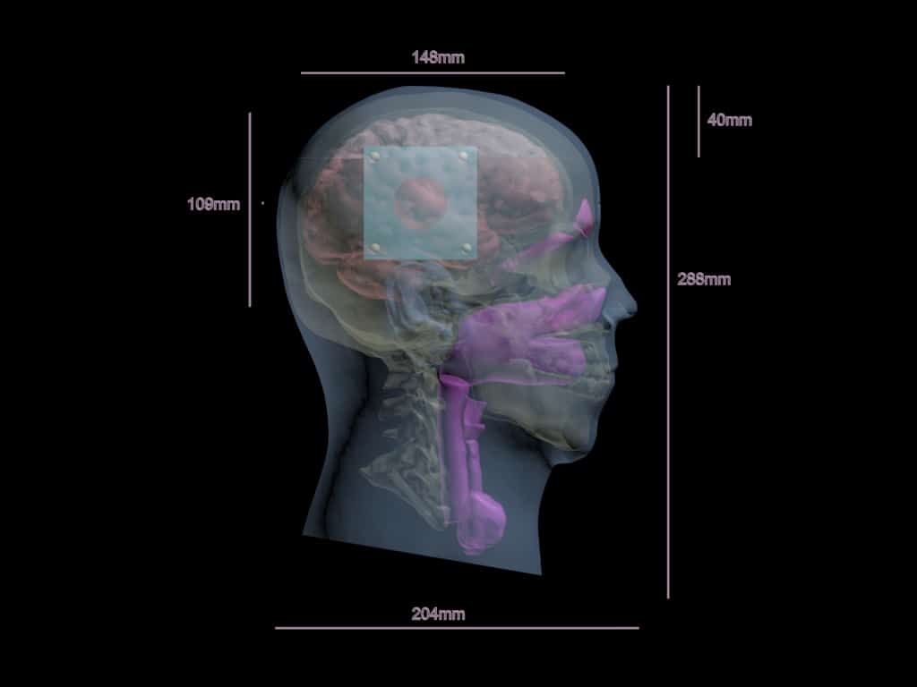
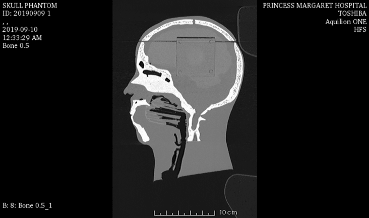
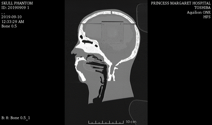
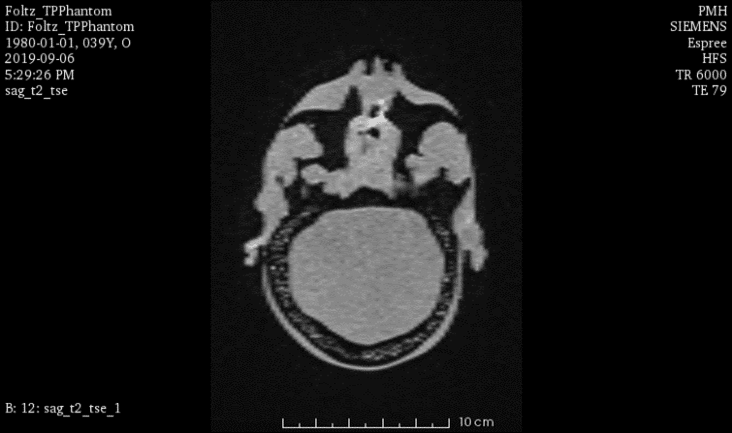
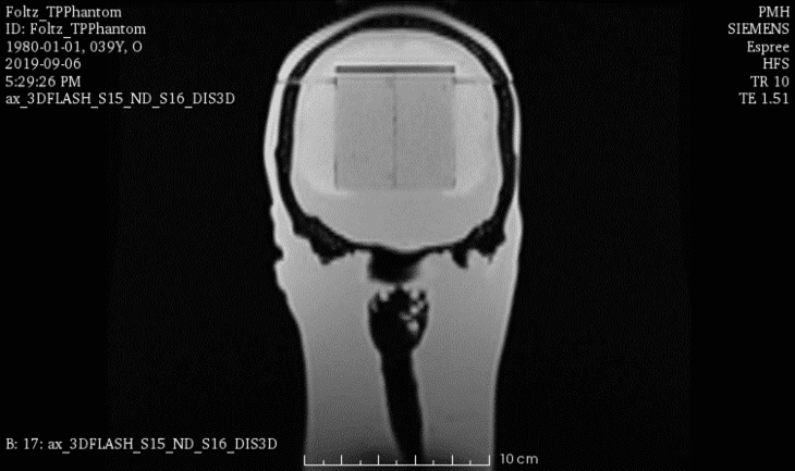
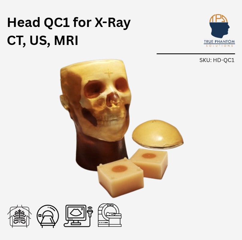
HD-QC1 is an anthropomorphic adult human head phantom that can be used for applications related to Image guided radiotherapy (IGRT), Stereotactic radiosurgery (SRS), and potential quality assurance (QA) applications. It serves as an ideal tool to study target localization and dose delivery under the image guidance of X-Ray/CT, US, MRI and PET.
Head Models: Dynamic
Appearance: Amber Translucent
This product can be customized based on your requirements — contact us.
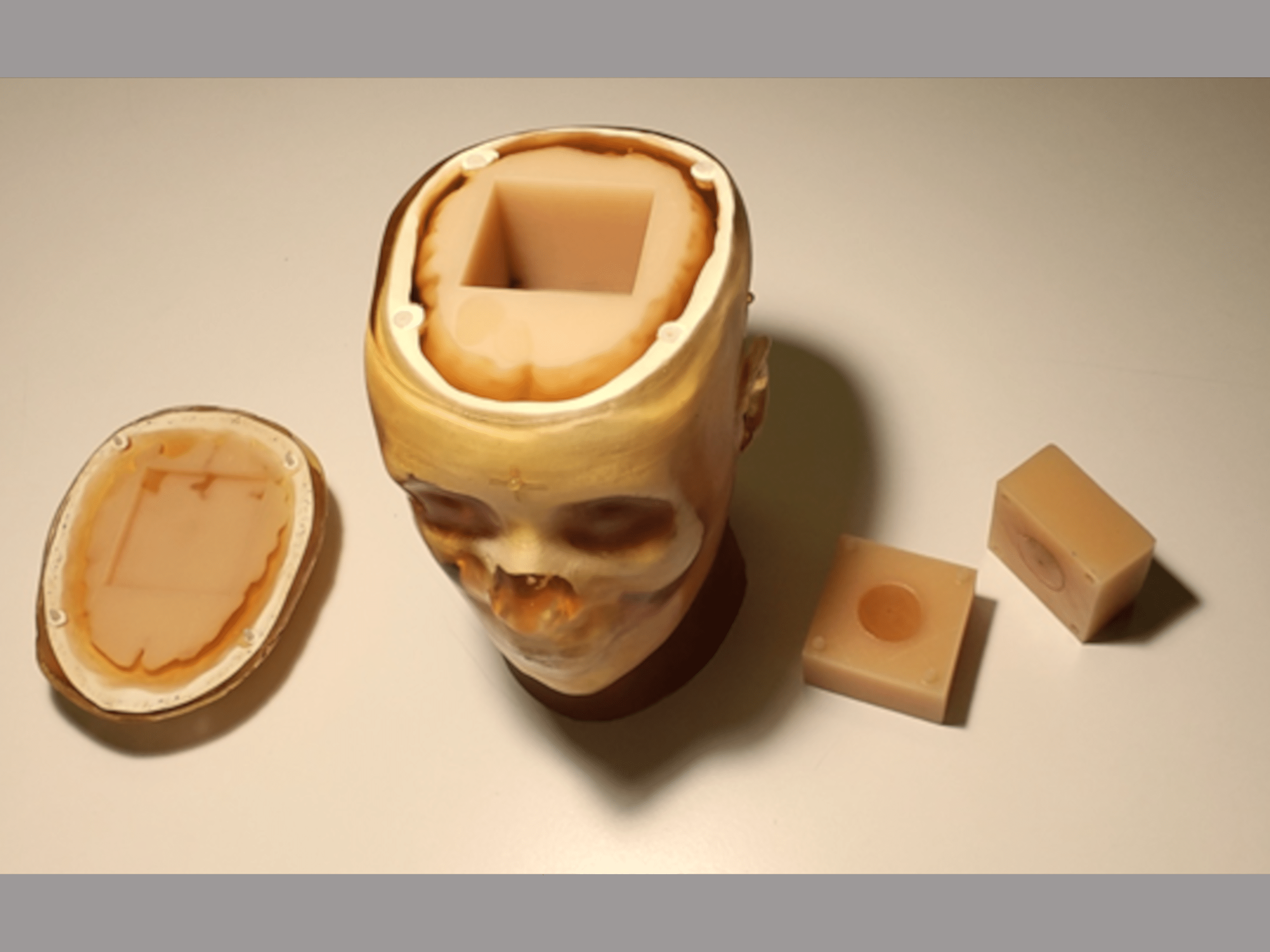
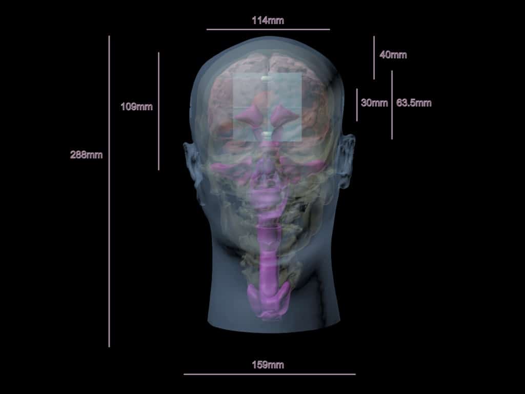
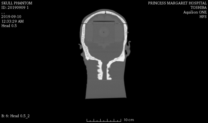
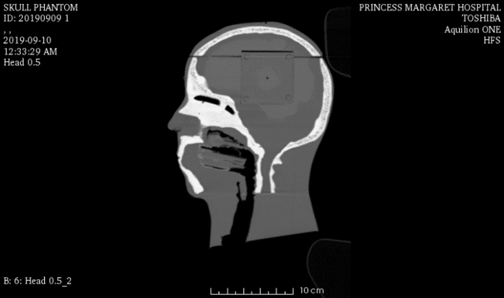
HD-QC1 serves as an ideal tool to study target localization and dose delivery under the image guidance of X-Ray/CT, US, MRI, and PET. The anthropomorphic features include a lifelike skull with cortical and trabecular bone structures, simulated brain(white and gray matter), cervical vertebrae, sinus cavities, larynx, trachea, and oral cavities.
In addition to this, it can be customized with tissue-equivalent inserts for QA applications.
| Features | Dynamic, Adv. Head | Static Head | Dynamic Head | Standard Head | Training, V1 Head | Hematoma Head | Openable Head |
|---|---|---|---|---|---|---|---|
| Skull | ✓ | ✓ | ✓ | ✓ | ✓ | ✓ | ✓ |
| Openable and Removable Calvaria | ✓ | ✖ | ✖ | ✖ | ✖ | ✖ | ✓ |
| Cervical Spine | ✖ | ✓ | ✖ | ✖ | ✓ | ✓ | ✖ |
| Skin Mimicking Material | ✓ | ✓ | ✓ | ✓ | ✓ | ✓ | ✓ |
| Brain (Gray Matter) | ✓ | ✓ | ✓ | ✓ | ✓ | ✓ | ✖ |
| Brain Ventricles | ✓ | ✖ | ✖ | ✓ | ✖ | ✓ | ✖ |
| Brain Tumor | ✖ | ✖ | ✖ | ✓ | ✖ | ✖ | ✖ |
| Falx Cerebri | ✖ | ✖ | ✖ | ✓ | ✓ | ✓ | ✖ |
| White Matter | ✖ | ✖ | ✖ | ✖ | ✓ | ✖ | ✖ |
| Brain Vessels | ✓ ( {2mm, 3mm, 4mm} incorporated Circle of Willis ) | ✖ | ✓ ( {2mm, 3mm, 4mm} incorporated Circle of Willis ) | ✓ | ✖ | ✖ | ✖ |
| One Bifurcation | ✓ | ✖ | ✓ | ✓ | ✖ | ✖ | ✖ |
| One Stenosis | ✓ | ✖ | ✓ | ✖ | ✖ | ✖ | ✖ |
| Two Aneurysms | ✓ | ✖ | ✓ | ✖ | ✖ | ✖ | ✖ |
| Vessels Connected with the Neck | ✓ | ✖ | ✓ | ✓ | ✖ | ✖ | ✖ |
| Plastic Connectors | ✓ | ✖ | ✓ | ✖ | ✖ | ✖ | ✖ |
| Small Static Objects | ✖ | ✓ ( {0.5mm, 1mm, 2mm, 3mm Diameters} ) | ✖ | ✖ | ✖ | ✖ | ✖ |
| Realistic Air Cavities | ✖ | ✖ | ✖ | ✖ | ✓ | ✖ | ✖ |
| Alignment Markings | ✓ | ✓ | ✓ | ✓ | ✓ | ✓ | ✓ |
| Frontal Sinus with Air Gaps | ✖ | ✖ | ✖ | ✖ | ✓ | ✖ | ✖ |
| Hematoma | ✖ | ✖ | ✖ | ✖ | ✖ | ✓ | ✖ |
| Features<\/th>\n | Dynamic,\nAdv. Head<\/th>\n | Static\nHead<\/th>\n | Dynamic\nHead<\/th>\n | Standard\u00a0 \u00a0 \u00a0Head<\/th>\n | Training,\nV1 Head<\/th>\n | Hematoma Head<\/th>\n | Openable Head<\/th>\n<\/tr>\n<\/thead>\n | ||||||||||||||||||||||||||||||||||||||||||||||||||||||||||||||||||||||||||||||||||||||||||||||||||||||||||||||||||||||||||||||||||||||||||||||||||||||||||||||||||||||||||||||||||||||||||||||||||||||||||||||||||||||||||||||||||||||||||
|---|---|---|---|---|---|---|---|---|---|---|---|---|---|---|---|---|---|---|---|---|---|---|---|---|---|---|---|---|---|---|---|---|---|---|---|---|---|---|---|---|---|---|---|---|---|---|---|---|---|---|---|---|---|---|---|---|---|---|---|---|---|---|---|---|---|---|---|---|---|---|---|---|---|---|---|---|---|---|---|---|---|---|---|---|---|---|---|---|---|---|---|---|---|---|---|---|---|---|---|---|---|---|---|---|---|---|---|---|---|---|---|---|---|---|---|---|---|---|---|---|---|---|---|---|---|---|---|---|---|---|---|---|---|---|---|---|---|---|---|---|---|---|---|---|---|---|---|---|---|---|---|---|---|---|---|---|---|---|---|---|---|---|---|---|---|---|---|---|---|---|---|---|---|---|---|---|---|---|---|---|---|---|---|---|---|---|---|---|---|---|---|---|---|---|---|---|---|---|---|---|---|---|---|---|---|---|---|---|---|---|---|---|---|---|---|---|---|---|---|---|---|---|---|---|---|---|---|---|---|---|---|---|---|---|---|---|---|---|---|---|---|
| Skull<\/td>\n | \u2713<\/strong><\/td>\n| \u2713<\/span><\/td>\n | \u2713<\/span><\/td>\n | \u2713<\/span><\/td>\n | \u2713<\/span><\/td>\n | \u2713<\/span><\/td>\n | \u2713<\/span><\/td>\n<\/tr>\n | Openable and Removable Calvaria<\/td>\n | \u2713<\/strong><\/td>\n | \u2716<\/td>\n | \u2716<\/td>\n | \u2716<\/td>\n | \u2716<\/td>\n | \u2716<\/td>\n | \u2713<\/span><\/td>\n<\/tr>\n | Cervical Spine<\/td>\n | \u2716<\/span><\/td>\n | \u2713<\/span><\/td>\n | \u2716<\/span><\/td>\n | \u2716<\/span><\/td>\n | \u2713<\/span><\/td>\n | \u00a0\u2713<\/strong><\/td>\n | \u2716<\/span><\/td>\n<\/tr>\n | Skin Mimicking Material<\/td>\n | \u2713<\/strong><\/td>\n | \u2713<\/span><\/td>\n | \u2713<\/span><\/td>\n | \u2713<\/span><\/td>\n | \u2713<\/span><\/td>\n | \u2713<\/span><\/td>\n | \u2713<\/span><\/td>\n<\/tr>\n | Brain\n(Gray Matter)<\/td>\n | \u2713<\/strong><\/td>\n | \u2713<\/span><\/td>\n | \u2713<\/strong><\/td>\n | \u2713<\/span><\/td>\n | \u2713<\/span><\/td>\n | \u2713<\/span><\/td>\n | \u2716<\/span><\/td>\n<\/tr>\n | Brain Ventricles<\/td>\n | \u2713<\/strong><\/td>\n | \u2716<\/span><\/td>\n | \u2716<\/span><\/td>\n | \u2713<\/span><\/td>\n | \u2716<\/span><\/td>\n | \u2713<\/span><\/td>\n | \u2716<\/td>\n<\/tr>\n | Brain Tumor<\/td>\n | \u2716<\/span><\/td>\n | \u2716<\/span><\/td>\n | \u2716<\/span><\/td>\n | \u2713<\/span><\/td>\n | \u2716<\/span><\/td>\n | \u2716<\/span><\/td>\n | \u2716<\/span><\/td>\n<\/tr>\n | Falx Cerebri<\/td>\n | \u2716<\/span><\/td>\n | \u2716<\/span><\/td>\n | \u2716<\/span><\/td>\n | \u2713<\/span><\/td>\n | \u2713<\/span><\/td>\n | \u2713<\/span><\/td>\n | \u2716<\/span><\/td>\n<\/tr>\n | White Matter<\/td>\n | \u2716<\/span><\/td>\n | \u2716<\/span><\/td>\n | \u2716<\/span><\/td>\n | \u2716<\/span><\/td>\n | \u2713<\/span><\/td>\n | \u2716<\/span><\/td>\n | \u2716<\/td>\n<\/tr>\n | Brain Vessels<\/td>\n | \u2713 (\u00a0<\/strong>{2mm, 3mm, 4mm} incorporated Circle of Willis\u00a0<\/span>)<\/strong>\n<\/strong><\/td>\n | \u2716<\/span><\/td>\n | \u2713\u00a0<\/strong>(\u00a0<\/span>{<\/span>2mm, 3mm, 4mm} incorporated Circle of Willis\u00a0<\/span>)<\/span>\n<\/strong><\/td>\n | \u2713<\/strong><\/td>\n | \u2716<\/span><\/td>\n | \u2716<\/td>\n | \u2716<\/td>\n<\/tr>\n | One Bifurcation<\/td>\n | \u2713<\/span><\/td>\n | \u2716<\/span><\/td>\n | \u2713<\/span><\/td>\n | \u2713<\/strong><\/td>\n | \u2716<\/span><\/td>\n | \u2716<\/span><\/td>\n | \u2716<\/td>\n<\/tr>\n | One Stenosis<\/td>\n | \u2713<\/span><\/td>\n | \u2716<\/span><\/td>\n | \u2713<\/span><\/td>\n | \u2716<\/span><\/td>\n | \u2716<\/span><\/td>\n | \u2716<\/span><\/td>\n | \u2716<\/td>\n<\/tr>\n | Two Aneurysms<\/td>\n | \u2713<\/strong><\/td>\n | \u2716<\/span><\/td>\n | \u2713<\/span><\/td>\n | \u2716<\/td>\n | \u2716<\/td>\n | \u2716<\/td>\n | \u2716<\/td>\n<\/tr>\n | Vessels Connected with the Neck<\/td>\n | \u2713<\/span><\/td>\n | \u2716<\/strong><\/td>\n | \u2713<\/span><\/td>\n | \u2713<\/span><\/td>\n | \u2716<\/span><\/td>\n | \u2716<\/td>\n | \u2716<\/td>\n<\/tr>\n | Plastic Connectors<\/td>\n | \u2713<\/span><\/td>\n | \u2716<\/td>\n | \u2713<\/span><\/td>\n | \u2716<\/td>\n | \u2716<\/td>\n | \u2716<\/td>\n | \u2716<\/td>\n<\/tr>\n | Small Static Objects<\/td>\n | \u2716<\/td>\n | \u2713 <\/span>(<\/strong> {0.5mm, 1mm, 2mm, 3mm Diameters} )<\/b><\/span><\/td>\n | \u2716<\/td>\n | \u2716<\/span><\/td>\n | \u2716<\/td>\n | \u2716<\/td>\n | \u2716<\/td>\n<\/tr>\n | Realistic Air Cavities<\/td>\n | \u2716<\/td>\n | \u2716<\/td>\n | \u2716<\/td>\n | \u2716<\/td>\n | \u2713<\/strong><\/td>\n | \u2716<\/td>\n | \u2716<\/td>\n<\/tr>\n | Alignment Markings<\/td>\n | \u2713<\/span><\/td>\n | \u2713<\/span><\/td>\n | \u2713<\/span><\/td>\n | \u2713<\/span><\/td>\n | \u2713<\/strong><\/td>\n | \u2713<\/span><\/td>\n | \u2713<\/span><\/td>\n<\/tr>\n | Frontal Sinus with Air Gaps<\/td>\n | \u2716<\/td>\n | \u2716<\/td>\n | \u2716<\/td>\n | \u2716<\/td>\n | \u2713<\/strong><\/td>\n | \u2716<\/td>\n | \u2716<\/td>\n<\/tr>\n | Hematoma<\/td>\n | \u2716<\/td>\n | \u2716<\/td>\n | \u2716<\/td>\n | \u2716<\/td>\n | \u2716<\/td>\n | \u2713<\/strong><\/td>\n | \u2716<\/td>\n<\/tr>\n<\/tbody>\n<\/table>"}}]}
| Technical Properties:
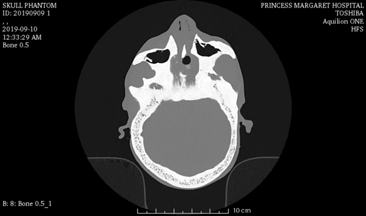
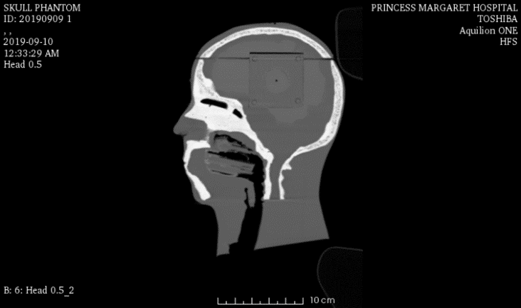
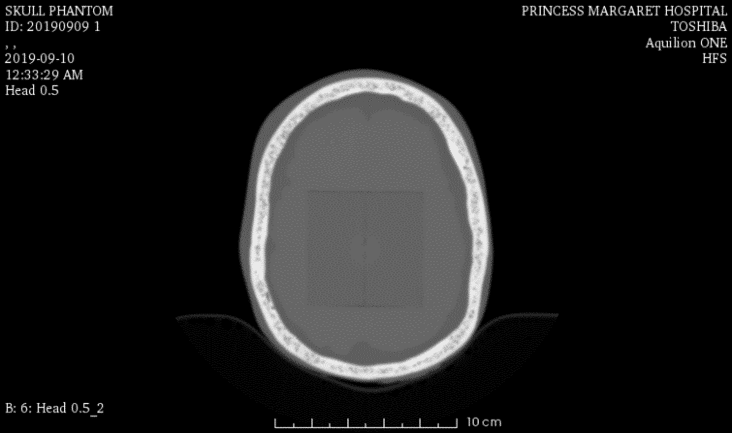
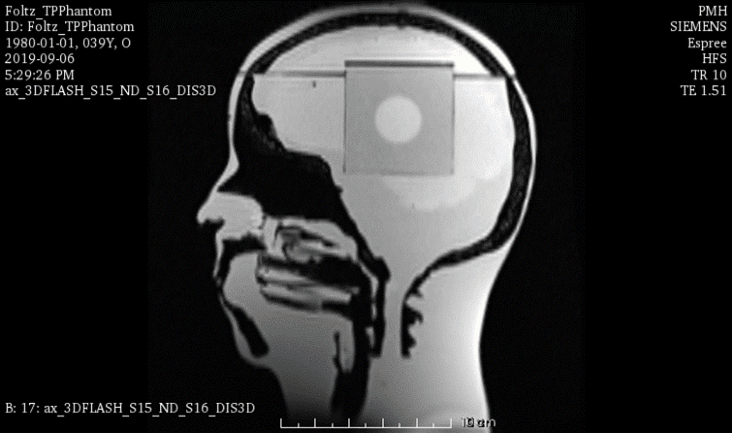
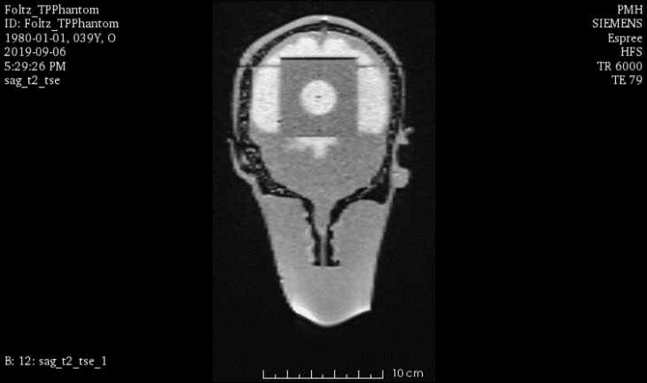
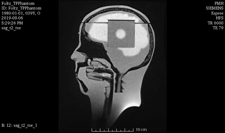
Sample scans of this phantom. Full scan galleries available on request. 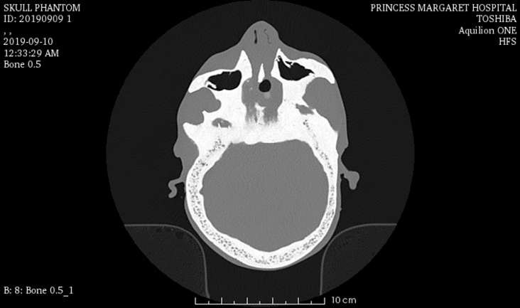
Similar products
| ||||||||||||||||||||||||||||||||||||||||||||||||||||||||||||||||||||||||||||||||||