X-Ray, CT, US, MRI Phantoms
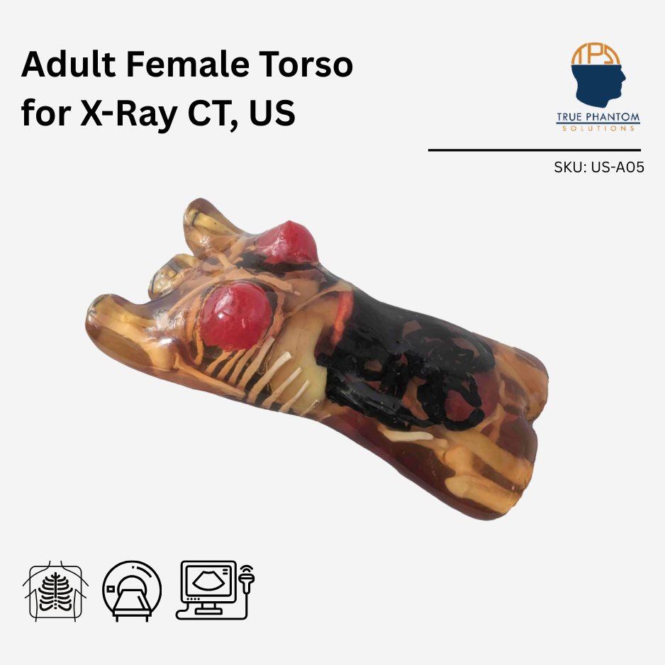
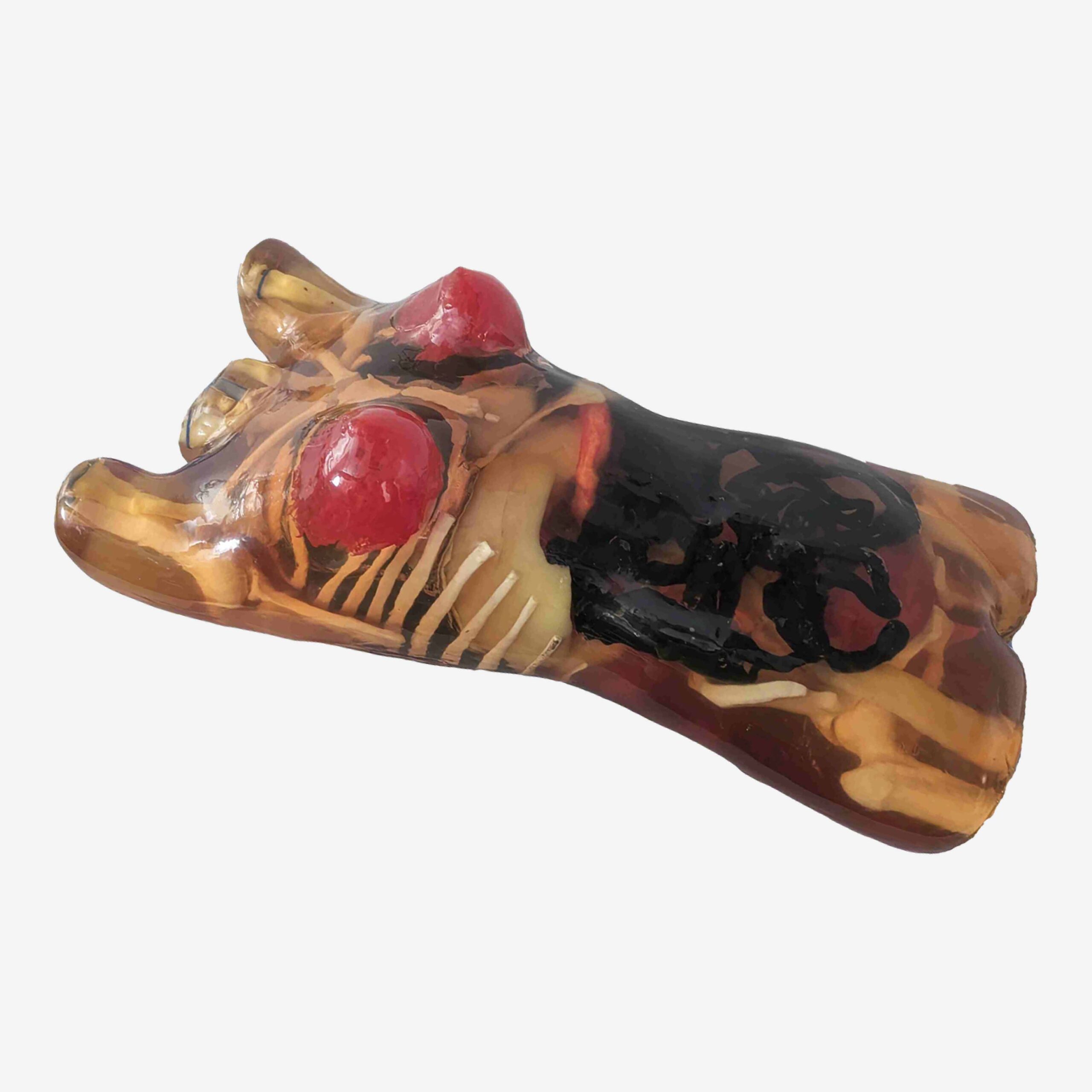
Adult Female Torso for X-Ray CT, US
Quote-based pricing
This phantom was designed using a CT dataset from a healthy 35-year-old female patient, standing 168 cm (5’5″) tall with a BMI adjusted to 22. The model was developed using advanced artificial intelligence (AI) software, incorporating a leading-edge algorithm to extract anatomical structures, ensuring exceptional precision and anatomical accuracy within the phantom.
Torso Model: Female (Dynamic)
Model: X-Ray CT
This product can be customized based on your requirements — contact us.
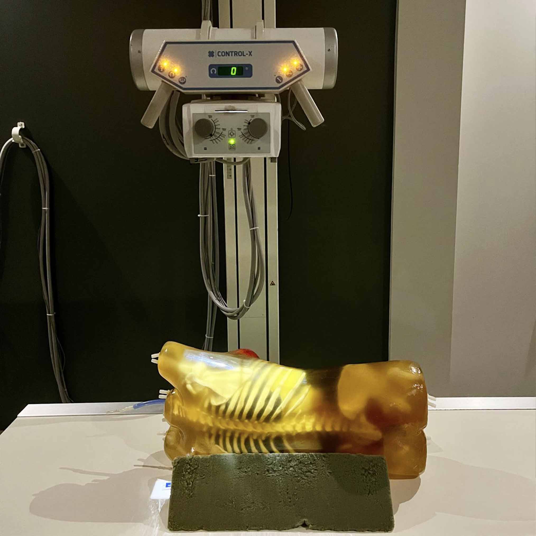
The Adult Female Torso Phantom is a groundbreaking innovation in medical imaging simulation, offering an unprecedented level of anatomical accuracy. With the help of AI (Artificial Intelligence) software, this phantoms design has been directly extracted from CT scans of a live, healthy 35-year-old patient with a height of 168cm (5’5″) and a BMI adjusted to 22. As a result, it features highly realistic anatomical structures and organ positioning. Developed in collaboration with medical professionals in the United States and Switzerland, this advanced phantom sets a new standard in medical imaging education, system development, and research applications.
Engineered with multi-modality compatibility, the Adult Female Torso Phantom is optimized for X-Ray, CT, and Ultrasound, making it a versatile tool for a wide range of clinical and research purposes. Its advanced tissue-mimicking materials closely replicate soft tissue and bone of real human tissue. This high level of realism is particularly beneficial for radiology training, where medical students, residents, and technologists can gain hands-on experience using this phantom with different imaging equipment.
This female torso phantom also offers insight into several specialties dedicated to women’s health, as well as general medical fields that frequently address female-specific conditions. For example, gynecology, breast oncology, and mammography, with the potential for customizations to address obstetrics, including childbirth and pregnancy.
Note: By default, the female torso phantom comes in a translucent amber appearance. The skin feature can be included upon request.
| Type of Tissue | Sound Velocity[m/s] | Density [g/cm3] | Attenuation Measured at 2.25 MHz [dB/cm] | Hardness [Shore OO] | T2 [ms] | Speckles |
| Organs with Speckles (liver, kidneys, etc.) | 1435 ± 10 | 0.99 | 1.0 ± 0.2 | 20 | 70 | VARIABLE |
| Organs without Speckles (stomach, intestines, etc.) | 1400 ± 10 | 0.99 | 1.0 ± 0.2 | 20 | 70 | NO |
| Body Tissue | 1410 ± 10 | 1.00 | 1.2 ± 0.2 | 30 | 65 | NO/LOW |
| Cortical Bone | 3000 ± 30 | 2.31 | 6.4 ± 0.3 | N/A | N/A | N/A |
| Trabecular Bone | 2800 ± 50 | 2.03 | 21 ± 2 | N/A | N/A | N/A |
| Thermal Properties of Bone-Mimicking Material | |||||
| Thermal Conductivity | Volumetric Specific Heat Capacity | Thermal Diffusivity | Thermal Resistivity | Specific Heat | Speed of Sound |
| 0.776 W/ m K | 1.040 MJ/ m^3 K | 0.746 mm^2/ s | 1.289 m K/ W | 0.978 J/ g Deg Celsius | 3070 m/s |
| Tissue Type | HU Value (Average) |
| Body Tissue | -25 |
| Trabecular Bone | 800 |
| Cortical Bone | 1300 |
| Aorta | 40 |
| Vena Cava | 40 |
| Trachea | 80 (Tissue Part), -1000 (Air Filled Part) |
| Lungs | -850 |
| Pancreas | 110 |
| Diaphragm | 80 |
| Mammary Fat Tissues | -25 |
| Mammary Gland Tissues | 35 |
| Cervix | 50 |
| Vaginal Canal | 50 |
| Tissue Type | HU Value (Average) |
| Spleen | 110 |
| Kidneys | 110 |
| Bladder | 35 |
| Rectum Wall | 100 |
| Sigmoid Colon Wall | 100 |
| Heart | 40 |
| Liver | 110 |
| Gallbladder | 35 |
| Stomach | -850 |
| Large and Small Intestines | 80 (Tissue Part), -1000 (Air Filled Part) |
| Ovaries | 50 |
| Uterus | 50 |
| Torso Model | Female (Dynamic), Female (Standard) |
| Model | X-Ray CT, US, X-Ray CT, MRI |
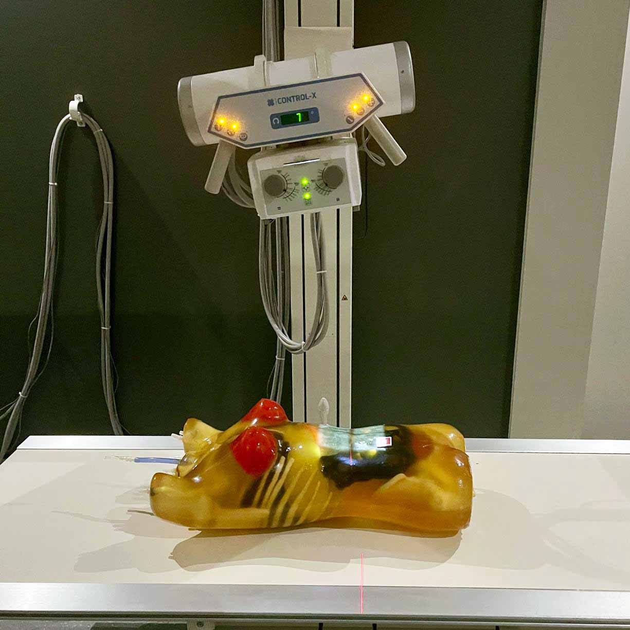
Sample scans of this phantom. Full scan galleries available on request.
Similar products