

X-Ray, CT, US, MRI Phantoms
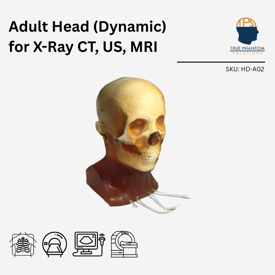
Adult Head (Dynamic) for X-Ray CT, US, MRI
$9,980
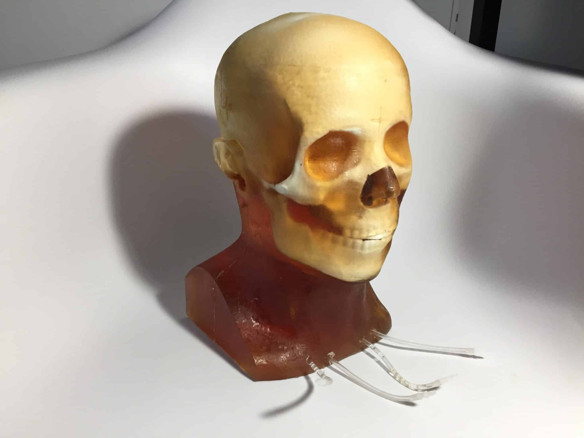
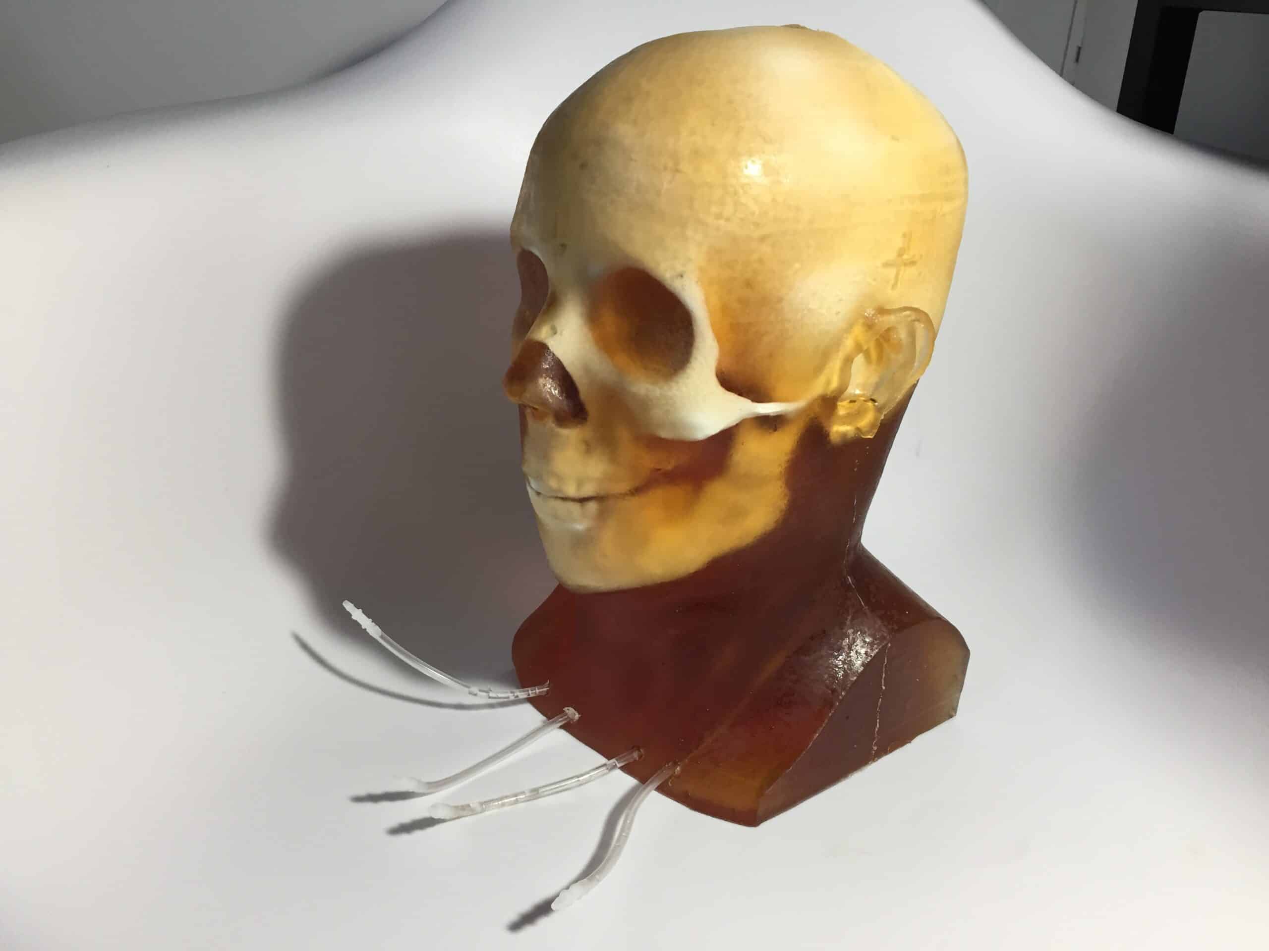
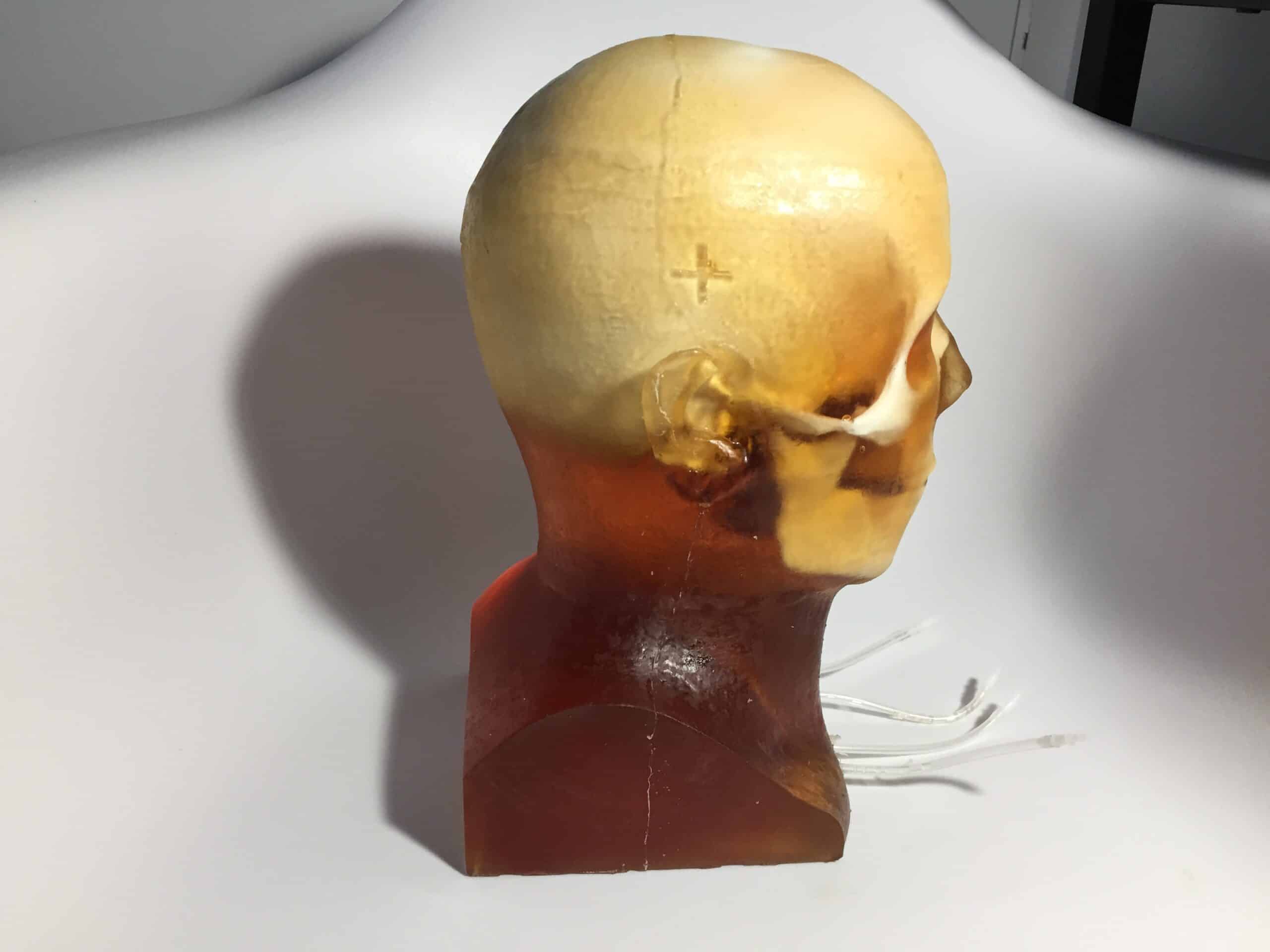
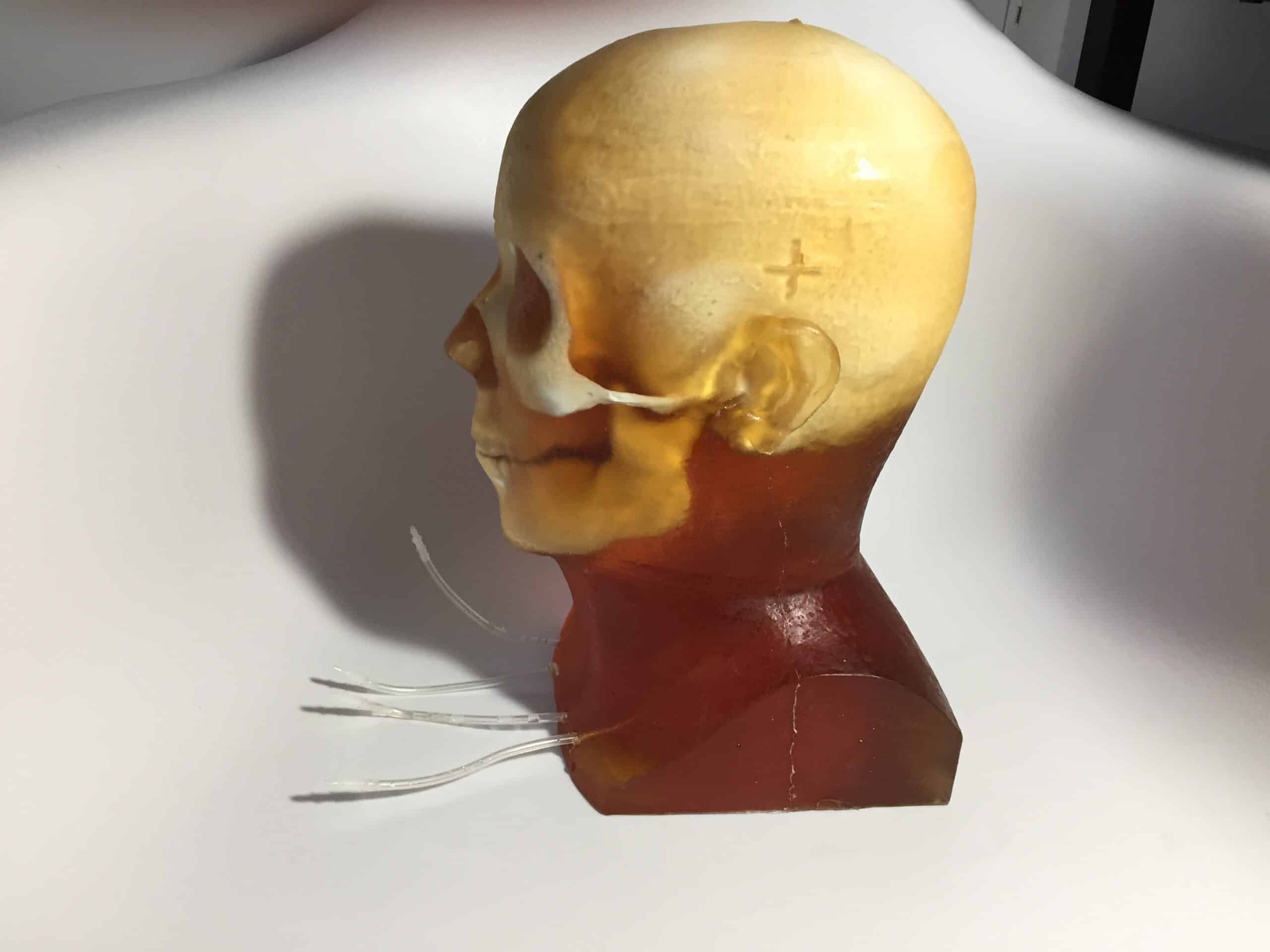
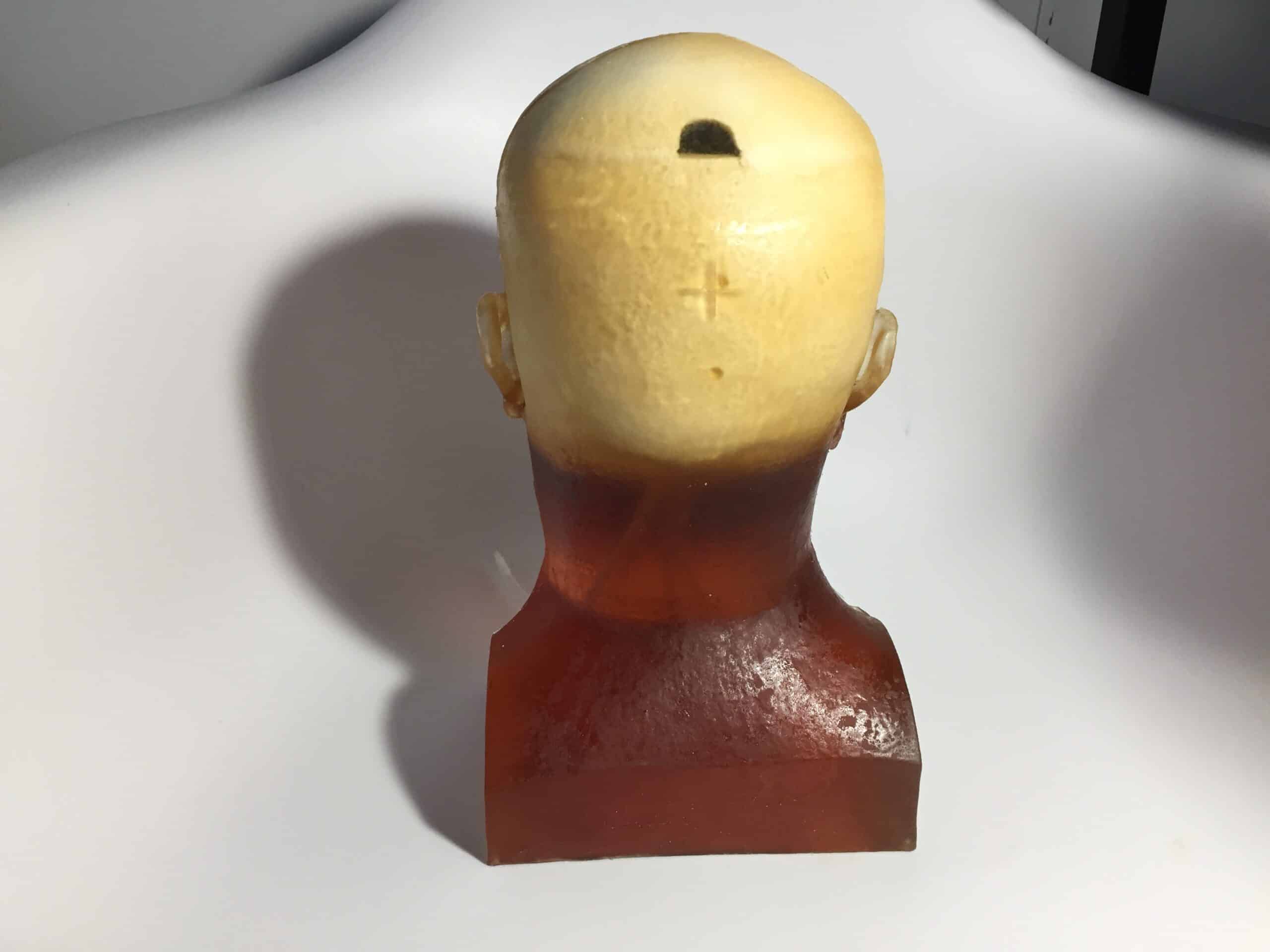
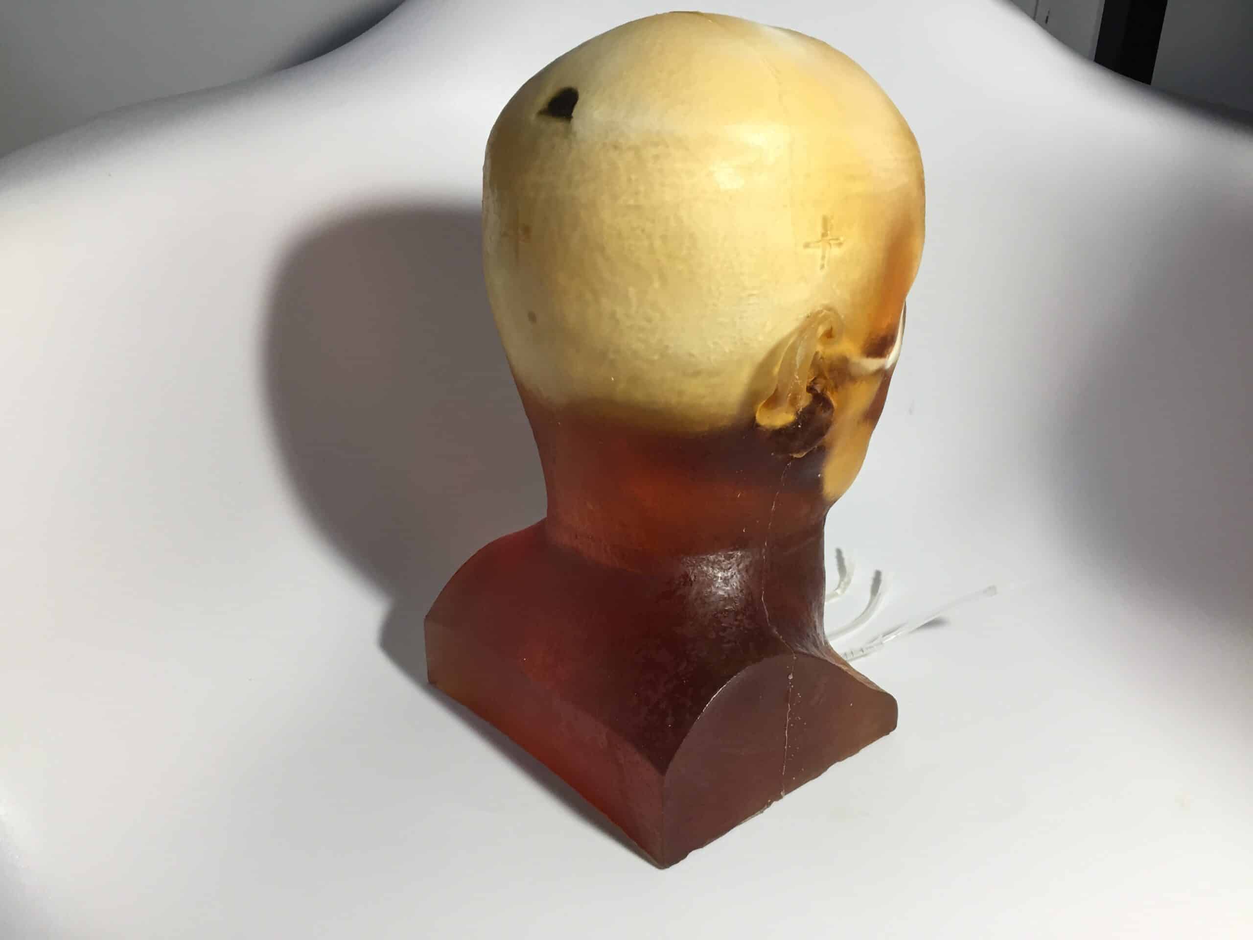
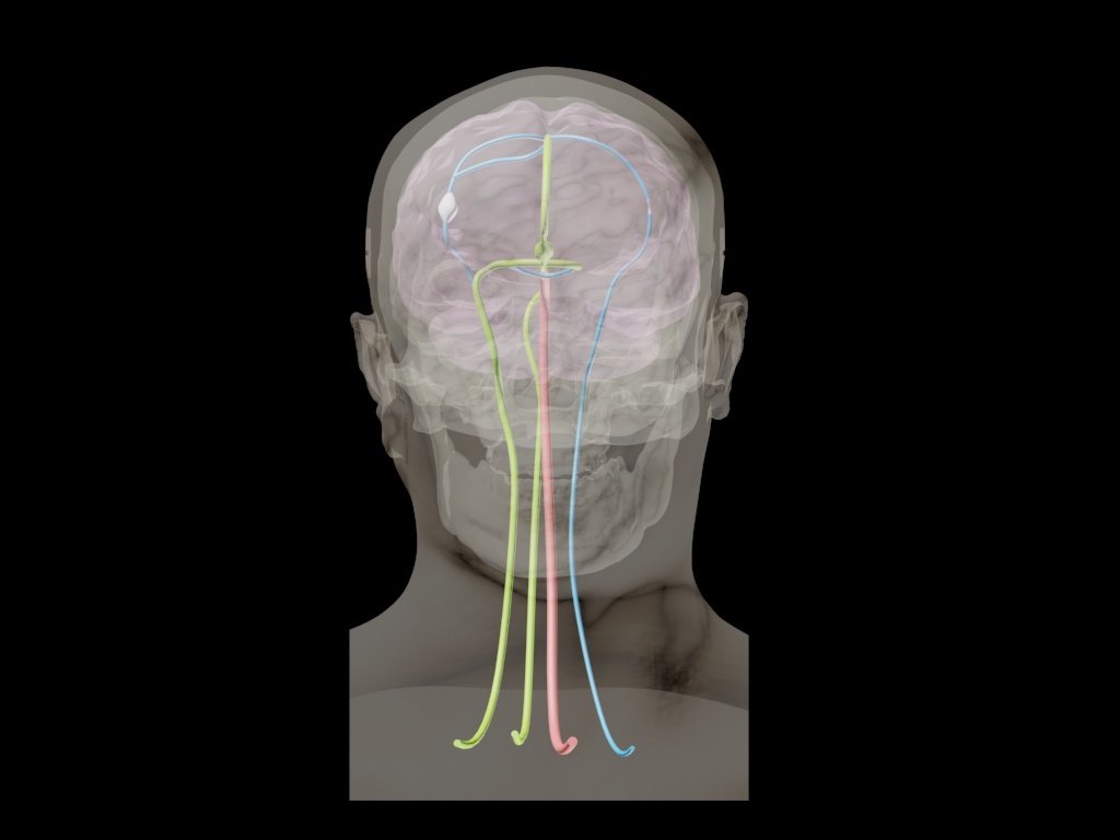
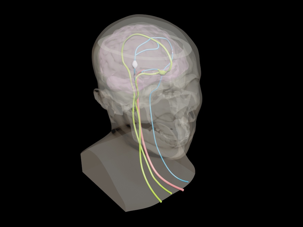
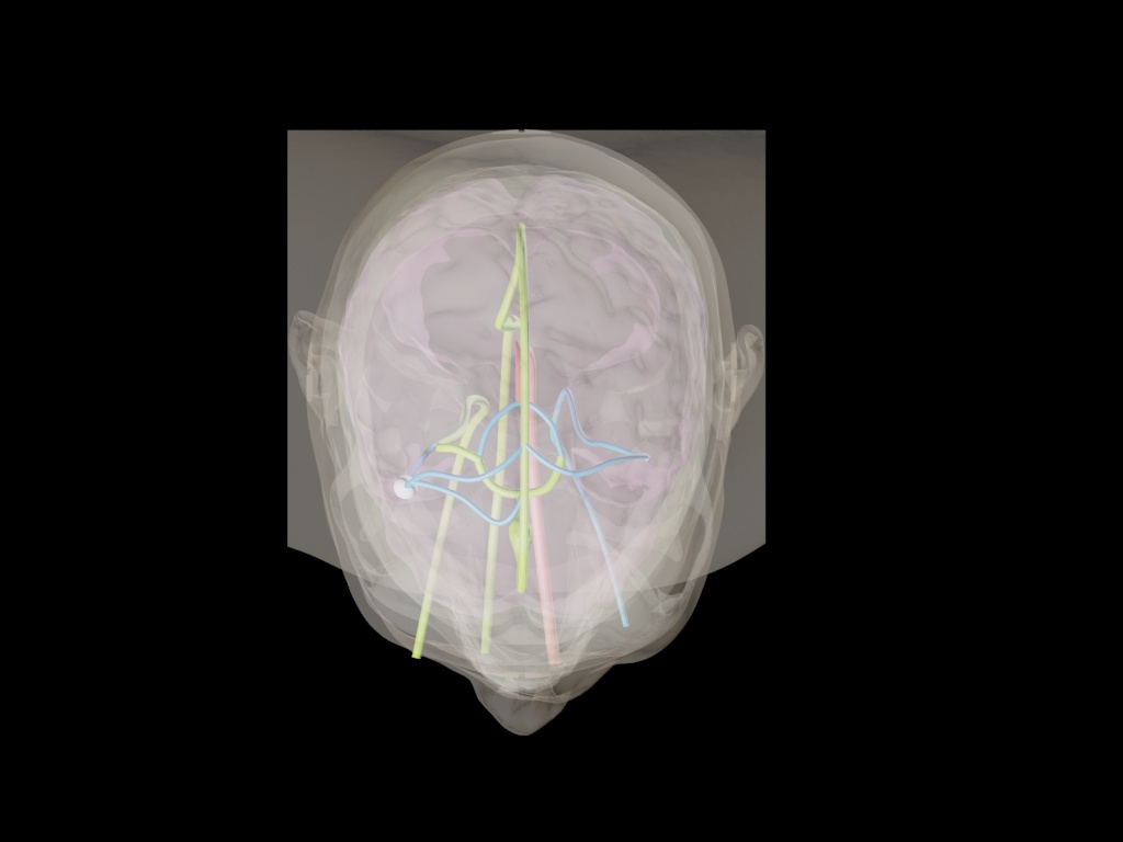
The Adult Head (Dynamic) Phantom is detectable on Ultrasound, MRI, and X-Ray/CT scans. Its brain parenchyma is made of an ultra-soft polyurethane-based material that resembles the soft tissue of a human brain. The skull bone features a realistic three-layered structure with an inner diploe layer. Additionally, the phantom includes realistic blood vessels that can be filled with commonly used blood-mimicking fluid, with or without a contrast agent.
Head Models: Dynamic
Appearance: Amber Translucent
This product can be customized based on your requirements — contact us.
The Adult Head (Dynamic) phantom is visible on Ultrasound, MRI, and X-Ray/CT scans. This head phantom can also be utilized for angiography training and applications. The brain parenchyma is made of an ultra-soft polyurethane-based material that mimics soft tissue, and its anatomical shape was created based on the MRI scan of a human brain. The materials used to build this phantom are stable with time, and they do not dry out. The skull bone has a realistic three-layered structure with an inner diploe layer. This phantom can be used with Barium-sulfate, Iodine-based, Gadolinium-based, Microbubble, and Iron Oxide-based contrast mediums.
In terms of MRI applications, the phantom tissues have realistic T2 relaxation time values, which makes this product the best fit for any T2-weighted MRI imaging methods. Very good results can also be achieved with Proton Density imaging methods. The phantom can be still imaged with T1-weighted methods, but the T1 values are less realistic, and they are within the range of about 100 ms.
The phantom comprises realistic blood vessels which can be filled with any commonly used blood-mimicking fluid with or without a contrast agent.
Note: For best results, prior to imaging the phantom, please fill the blood vessels with water or chosen contrast agent.
| Features | Dynamic, Adv. Head | Static Head | Dynamic Head | Standard Head | Training, V1 Head | Hematoma Head | Openable Head |
|---|---|---|---|---|---|---|---|
| Skull | ✓ | ✓ | ✓ | ✓ | ✓ | ✓ | ✓ |
| Openable and Removable Calvaria | ✓ | ✖ | ✖ | ✖ | ✖ | ✖ | ✓ |
| Cervical Spine | ✖ | ✓ | ✖ | ✖ | ✓ | ✓ | ✖ |
| Skin Mimicking Material | ✓ | ✓ | ✓ | ✓ | ✓ | ✓ | ✓ |
| Brain (Gray Matter) | ✓ | ✓ | ✓ | ✓ | ✓ | ✓ | ✖ |
| Brain Ventricles | ✓ | ✖ | ✖ | ✓ | ✖ | ✓ | ✖ |
| Brain Tumor | ✖ | ✖ | ✖ | ✓ | ✖ | ✖ | ✖ |
| Falx Cerebri | ✖ | ✖ | ✖ | ✓ | ✓ | ✓ | ✖ |
| White Matter | ✖ | ✖ | ✖ | ✖ | ✓ | ✖ | ✖ |
| Brain Vessels | ✓ ( {2mm, 3mm, 4mm} incorporated Circle of Willis ) | ✖ | ✓ ( {2mm, 3mm, 4mm} incorporated Circle of Willis ) | ✓ | ✖ | ✖ | ✖ |
| One Bifurcation | ✓ | ✖ | ✓ | ✓ | ✖ | ✖ | ✖ |
| One Stenosis | ✓ | ✖ | ✓ | ✖ | ✖ | ✖ | ✖ |
| Two Aneurysms | ✓ | ✖ | ✓ | ✖ | ✖ | ✖ | ✖ |
| Vessels Connected with the Neck | ✓ | ✖ | ✓ | ✓ | ✖ | ✖ | ✖ |
| Plastic Connectors | ✓ | ✖ | ✓ | ✖ | ✖ | ✖ | ✖ |
| Small Static Objects | ✖ | ✓ ( {0.5mm, 1mm, 2mm, 3mm Diameters} ) | ✖ | ✖ | ✖ | ✖ | ✖ |
| Realistic Air Cavities | ✖ | ✖ | ✖ | ✖ | ✓ | ✖ | ✖ |
| Alignment Markings | ✓ | ✓ | ✓ | ✓ | ✓ | ✓ | ✓ |
| Frontal Sinus with Air Gaps | ✖ | ✖ | ✖ | ✖ | ✓ | ✖ | ✖ |
| Hematoma | ✖ | ✖ | ✖ | ✖ | ✖ | ✓ | ✖ |
| Features<\/th>\n | Dynamic,\nAdv. Head<\/th>\n | Static\nHead<\/th>\n | Dynamic\nHead<\/th>\n | Standard\u00a0 \u00a0 \u00a0Head<\/th>\n | Training,\nV1 Head<\/th>\n | Hematoma Head<\/th>\n | Openable Head<\/th>\n<\/tr>\n<\/thead>\n | ||||||||||||||||||||||||||||||||||||||||||||||||||||||||||||||||||||||||||||||||||||||||||||||||||||||||||||||||||||||||||||||||||||||||||||||||||||||||||||||||||||||||||||||||||||||||||||||||||||||||||||||||||||||||||||||||||||||||||
|---|---|---|---|---|---|---|---|---|---|---|---|---|---|---|---|---|---|---|---|---|---|---|---|---|---|---|---|---|---|---|---|---|---|---|---|---|---|---|---|---|---|---|---|---|---|---|---|---|---|---|---|---|---|---|---|---|---|---|---|---|---|---|---|---|---|---|---|---|---|---|---|---|---|---|---|---|---|---|---|---|---|---|---|---|---|---|---|---|---|---|---|---|---|---|---|---|---|---|---|---|---|---|---|---|---|---|---|---|---|---|---|---|---|---|---|---|---|---|---|---|---|---|---|---|---|---|---|---|---|---|---|---|---|---|---|---|---|---|---|---|---|---|---|---|---|---|---|---|---|---|---|---|---|---|---|---|---|---|---|---|---|---|---|---|---|---|---|---|---|---|---|---|---|---|---|---|---|---|---|---|---|---|---|---|---|---|---|---|---|---|---|---|---|---|---|---|---|---|---|---|---|---|---|---|---|---|---|---|---|---|---|---|---|---|---|---|---|---|---|---|---|---|---|---|---|---|---|---|---|---|---|---|---|---|---|---|---|---|---|---|---|
| Skull<\/td>\n | \u2713<\/strong><\/td>\n| \u2713<\/span><\/td>\n | \u2713<\/span><\/td>\n | \u2713<\/span><\/td>\n | \u2713<\/span><\/td>\n | \u2713<\/span><\/td>\n | \u2713<\/span><\/td>\n<\/tr>\n | Openable and Removable Calvaria<\/td>\n | \u2713<\/strong><\/td>\n | \u2716<\/td>\n | \u2716<\/td>\n | \u2716<\/td>\n | \u2716<\/td>\n | \u2716<\/td>\n | \u2713<\/span><\/td>\n<\/tr>\n | Cervical Spine<\/td>\n | \u2716<\/span><\/td>\n | \u2713<\/span><\/td>\n | \u2716<\/span><\/td>\n | \u2716<\/span><\/td>\n | \u2713<\/span><\/td>\n | \u00a0\u2713<\/strong><\/td>\n | \u2716<\/span><\/td>\n<\/tr>\n | Skin Mimicking Material<\/td>\n | \u2713<\/strong><\/td>\n | \u2713<\/span><\/td>\n | \u2713<\/span><\/td>\n | \u2713<\/span><\/td>\n | \u2713<\/span><\/td>\n | \u2713<\/span><\/td>\n | \u2713<\/span><\/td>\n<\/tr>\n | Brain\n(Gray Matter)<\/td>\n | \u2713<\/strong><\/td>\n | \u2713<\/span><\/td>\n | \u2713<\/strong><\/td>\n | \u2713<\/span><\/td>\n | \u2713<\/span><\/td>\n | \u2713<\/span><\/td>\n | \u2716<\/span><\/td>\n<\/tr>\n | Brain Ventricles<\/td>\n | \u2713<\/strong><\/td>\n | \u2716<\/span><\/td>\n | \u2716<\/span><\/td>\n | \u2713<\/span><\/td>\n | \u2716<\/span><\/td>\n | \u2713<\/span><\/td>\n | \u2716<\/td>\n<\/tr>\n | Brain Tumor<\/td>\n | \u2716<\/span><\/td>\n | \u2716<\/span><\/td>\n | \u2716<\/span><\/td>\n | \u2713<\/span><\/td>\n | \u2716<\/span><\/td>\n | \u2716<\/span><\/td>\n | \u2716<\/span><\/td>\n<\/tr>\n | Falx Cerebri<\/td>\n | \u2716<\/span><\/td>\n | \u2716<\/span><\/td>\n | \u2716<\/span><\/td>\n | \u2713<\/span><\/td>\n | \u2713<\/span><\/td>\n | \u2713<\/span><\/td>\n | \u2716<\/span><\/td>\n<\/tr>\n | White Matter<\/td>\n | \u2716<\/span><\/td>\n | \u2716<\/span><\/td>\n | \u2716<\/span><\/td>\n | \u2716<\/span><\/td>\n | \u2713<\/span><\/td>\n | \u2716<\/span><\/td>\n | \u2716<\/td>\n<\/tr>\n | Brain Vessels<\/td>\n | \u2713 (\u00a0<\/strong>{2mm, 3mm, 4mm} incorporated Circle of Willis\u00a0<\/span>)<\/strong>\n<\/strong><\/td>\n | \u2716<\/span><\/td>\n | \u2713\u00a0<\/strong>(\u00a0<\/span>{<\/span>2mm, 3mm, 4mm} incorporated Circle of Willis\u00a0<\/span>)<\/span>\n<\/strong><\/td>\n | \u2713<\/strong><\/td>\n | \u2716<\/span><\/td>\n | \u2716<\/td>\n | \u2716<\/td>\n<\/tr>\n | One Bifurcation<\/td>\n | \u2713<\/span><\/td>\n | \u2716<\/span><\/td>\n | \u2713<\/span><\/td>\n | \u2713<\/strong><\/td>\n | \u2716<\/span><\/td>\n | \u2716<\/span><\/td>\n | \u2716<\/td>\n<\/tr>\n | One Stenosis<\/td>\n | \u2713<\/span><\/td>\n | \u2716<\/span><\/td>\n | \u2713<\/span><\/td>\n | \u2716<\/span><\/td>\n | \u2716<\/span><\/td>\n | \u2716<\/span><\/td>\n | \u2716<\/td>\n<\/tr>\n | Two Aneurysms<\/td>\n | \u2713<\/strong><\/td>\n | \u2716<\/span><\/td>\n | \u2713<\/span><\/td>\n | \u2716<\/td>\n | \u2716<\/td>\n | \u2716<\/td>\n | \u2716<\/td>\n<\/tr>\n | Vessels Connected with the Neck<\/td>\n | \u2713<\/span><\/td>\n | \u2716<\/strong><\/td>\n | \u2713<\/span><\/td>\n | \u2713<\/span><\/td>\n | \u2716<\/span><\/td>\n | \u2716<\/td>\n | \u2716<\/td>\n<\/tr>\n | Plastic Connectors<\/td>\n | \u2713<\/span><\/td>\n | \u2716<\/td>\n | \u2713<\/span><\/td>\n | \u2716<\/td>\n | \u2716<\/td>\n | \u2716<\/td>\n | \u2716<\/td>\n<\/tr>\n | Small Static Objects<\/td>\n | \u2716<\/td>\n | \u2713 <\/span>(<\/strong> {0.5mm, 1mm, 2mm, 3mm Diameters} )<\/b><\/span><\/td>\n | \u2716<\/td>\n | \u2716<\/span><\/td>\n | \u2716<\/td>\n | \u2716<\/td>\n | \u2716<\/td>\n<\/tr>\n | Realistic Air Cavities<\/td>\n | \u2716<\/td>\n | \u2716<\/td>\n | \u2716<\/td>\n | \u2716<\/td>\n | \u2713<\/strong><\/td>\n | \u2716<\/td>\n | \u2716<\/td>\n<\/tr>\n | Alignment Markings<\/td>\n | \u2713<\/span><\/td>\n | \u2713<\/span><\/td>\n | \u2713<\/span><\/td>\n | \u2713<\/span><\/td>\n | \u2713<\/strong><\/td>\n | \u2713<\/span><\/td>\n | \u2713<\/span><\/td>\n<\/tr>\n | Frontal Sinus with Air Gaps<\/td>\n | \u2716<\/td>\n | \u2716<\/td>\n | \u2716<\/td>\n | \u2716<\/td>\n | \u2713<\/strong><\/td>\n | \u2716<\/td>\n | \u2716<\/td>\n<\/tr>\n | Hematoma<\/td>\n | \u2716<\/td>\n | \u2716<\/td>\n | \u2716<\/td>\n | \u2716<\/td>\n | \u2716<\/td>\n | \u2713<\/strong><\/td>\n | \u2716<\/td>\n<\/tr>\n<\/tbody>\n<\/table>\n<\/div>"}}]}
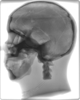
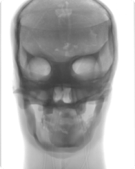
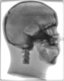
| X-Ray ScansNote: These scans are from a CUSTOM order of the phantom. The standard phantom does not include a cervical spine. It can be included upon request. For more imaging quality references, please see our Radiographs page. Technical Properties:
Similar products
| ||||||||||||||||||||||||||||||||||||||||||||||||||||||||||||||||||||||||||||||||||